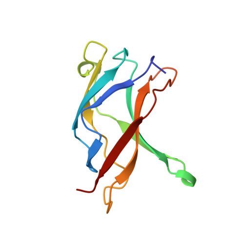
The role of DNA in the mechanism of NFkappaB dimer formation: crystal structures of the dimerization domains of the p50 and p65 subunits.
Huang, D.B., Huxford, T., Chen, Y.Q., Ghosh, G.(1997) Structure 5: 1427-1436
- PubMed: 9384558 Search on PubMed
- DOI: https://doi.org/10.1016/s0969-2126(97)00293-1
- Primary Citation Related Structures:
1BFS, 1BFT - PubMed Abstract:
Members of the rel/NFkappaB family of transcription factors play a vital role in the regulation of rapid cellular responses, such as those required to fight infection or react to cellular stress. Members of this family of proteins form homo- and heterodimers with differing affinities for dimerization. They share a structural motif known as the rel homology region (RHR), the C-terminal one third of which mediates protein dimerization. Crystal structures of the rel/NFkappaB family members p50 and p65 in their DNA-bound homodimeric form have been solved. These structures showed that the residues from the dimerization domains of both p50 and p65 participate in DNA binding and that the DNA-protein and protein dimerization surfaces form one continuous overlapping interface. We desired to investigate the contribution of DNA to NFkappaB dimerization and to identify the mechanism for the selective association of rel/NFkappaB family peptides into transcriptionally active dimers. We report here the crystal structures of the dimerization domains of murine p50 and p65 at 2.2 A and 2.0 A resolution, respectively. A comparison of these two structures suggests that conservative amino acid changes at three positions are responsible for the differences in their dimer interfaces. The presence of the target DNA does not change the dimer interface of either protein in any significant manner. These two structures suggest that the rel/NFkappaB family of transcription factors use only a few conservative changes in their amino acid sequences to form a host of dimers with varying affinities for dimerization. Amino acids at positions corresponding to 254, 267, and 307 of murine p50, function as primary determinants for the observed differences in dimerization affinity. The DNA-contacting charged amino acid sidechains from the dimerization domains are held in a similar conformation in both the DNA-bound and free states, therefore, no major structural rearrangement is required to bring these residues into contact with the DNA.
- Department of Chemistry and Biochemistry, University of California, San Diego, 9500 Gilman Drive, La Jolla, CA 92093-0359, USA.
Organizational Affiliation: