1BF3

P-HYDROXYBENZOATE HYDROXYLASE (PHBH) MUTANT WITH CYS 116 REPLACED BY SER (C116S) AND ARG 42 REPLACED BY LYS (R42K), IN COMPLEX WITH FAD AND 4-HYDROXYBENZOIC ACID


Experimental Data Snapshot

  • Method: X-RAY DIFFRACTION
  • Resolution: 2.20 Å
  • R-Value Work: 0.175 

wwPDB Validation   3D Report Full Report


This is version 1.8 of the entry. See complete history


Literature

Lys42 and Ser42 variants of p-hydroxybenzoate hydroxylase from Pseudomonas fluorescens reveal that Arg42 is essential for NADPH binding.

Eppink, M.H.Schreuder, H.A.van Berkel, W.J.

(1998) Eur J Biochem 253: 194-201

  • DOI: https://doi.org/10.1046/j.1432-1327.1998.2530194.x
  • Primary Citation of Related Structures:  
    1BF3

  • PubMed Abstract: 

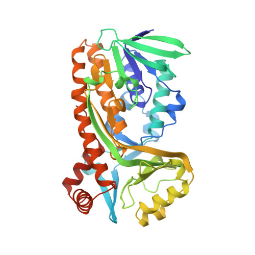
    The conserved Arg42 of the flavoprotein p-hydroxybenzoate hydroxylase is located at the entrance of the active site in a loop between helix H2 and sheet E1 of the FAD-binding domain. Replacement of Arg42 by Lys or Ser decreases the turnover rate of p-hydroxybenzoate hydroxylase from Pseudomonas fluorescens by more than two orders of magnitude. Rapid reaction kinetics show that the low activity of the Arg42 variants results from impaired binding of NADPH. In contrast to an earlier conclusion drawn for p-hydroxybenzoate hydroxylase from Acinetobacter calcoaceticus, substitution of Arg42 with Ser42 in the enzyme from P. fluorescens hardly disturbs the binding of FAD. Crystals of [Lys42]p-hydroxybenzoate hydroxylase complexed with 4-hydroxybenzoate diffract to 0.22-nm resolution. The structure of the Lys42 variant is virtually indistinguishable from the native enzyme with the flavin ring occupying the interior position within the active site. Lys42 in the mutant structure interacts indirectly via a solvent molecule with the 3-OH of the adenosine ribose moiety of FAD. Substrate perturbation difference spectra suggest that the Arg42 replacements influence the solvent accessibility of the flavin ring in the oxidized enzyme. In spite of this, the Arg42 variants fully couple enzyme reduction to substrate hydroxylation. Sequence-comparison studies suggest that Arg42 is involved in binding of the 2'-phosphoadenosine moiety of NADPH.


  • Organizational Affiliation

    Department of Biomolecular Sciences, Wageningen Agricultural University, The Netherlands.


Macromolecules
Find similar proteins by:  (by identity cutoff)  |  3D Structure
Entity ID: 1
MoleculeChains Sequence LengthOrganismDetailsImage
P-HYDROXYBENZOATE HYDROXYLASE394Pseudomonas fluorescensMutation(s): 2 
Gene Names: POBA
EC: 1.14.13.2
UniProt
Find proteins for P00438 (Pseudomonas fluorescens)
Explore P00438 
Go to UniProtKB:  P00438
Entity Groups  
Sequence Clusters30% Identity50% Identity70% Identity90% Identity95% Identity100% Identity
UniProt GroupP00438
Sequence Annotations
Expand
  • Reference Sequence
Small Molecules
Ligands 2 Unique
IDChains Name / Formula / InChI Key2D Diagram3D Interactions
FAD
Query on FAD

Download Ideal Coordinates CCD File 
B [auth A]FLAVIN-ADENINE DINUCLEOTIDE
C27 H33 N9 O15 P2
VWWQXMAJTJZDQX-UYBVJOGSSA-N
PHB
Query on PHB

Download Ideal Coordinates CCD File 
C [auth A]P-HYDROXYBENZOIC ACID
C7 H6 O3
FJKROLUGYXJWQN-UHFFFAOYSA-N
Binding Affinity Annotations 
IDSourceBinding Affinity
PHB Binding MOAD:  1BF3 Kd: 1.20e+5 (nM) from 1 assay(s)
Experimental Data & Validation

Experimental Data

  • Method: X-RAY DIFFRACTION
  • Resolution: 2.20 Å
  • R-Value Work: 0.175 
  • Space Group: C 2 2 21
Unit Cell:
Length ( Å )Angle ( ˚ )
a = 71.35α = 90
b = 145.9β = 90
c = 88.8γ = 90
Software Package:
Software NamePurpose
X-PLORmodel building
X-PLORrefinement
XDSdata reduction
XSCALEdata scaling
X-PLORphasing

Structure Validation

View Full Validation Report


Entry History 

Deposition Data

Revision History  (Full details and data files)

  • Version 1.0: 1998-08-12
    Type: Initial release
  • Version 1.1: 2008-03-24
    Changes: Version format compliance
  • Version 1.2: 2011-07-13
    Changes: Derived calculations, Version format compliance
  • Version 1.3: 2014-02-12
    Changes: Refinement description
  • Version 1.4: 2014-04-09
    Changes: Data collection
  • Version 1.5: 2017-11-29
    Changes: Derived calculations, Other
  • Version 1.6: 2018-04-04
    Changes: Data collection
  • Version 1.7: 2021-11-03
    Changes: Database references, Derived calculations
  • Version 1.8: 2023-08-02
    Changes: Refinement description