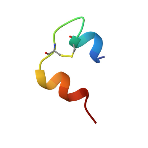
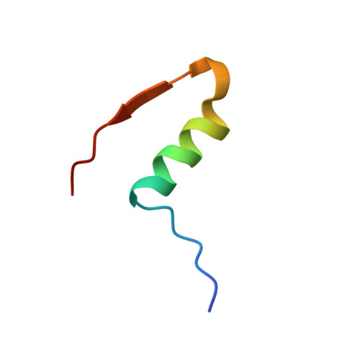
Structure of an insulin dimer in an orthorhombic crystal: the structure analysis of a human insulin mutant (B9 Ser-->Glu).
Yao, Z.P., Zeng, Z.H., Li, H.M., Zhang, Y., Feng, Y.M., Wang, D.C.(1999) Acta Crystallogr D Biol Crystallogr 55: 1524-1532
- PubMed: 10489447 Search on PubMed
- DOI: https://doi.org/10.1107/s0907444999008562
- Primary Citation Related Structures:
1B9E - PubMed Abstract:
The structure of human insulin mutant B9 (Ser-->Glu) was determined by an X-ray crystallographic method at 2.5 A resolution with an R factor of 0.165 under non-crystallographic restraints. The crystals were grown at low pH (<3.8) and belong to the orthorhombic P2(1)2(1)2(1) space group with unit-cell dimensions a = 44.54, b = 46.40, c = 51.85 A and one dimer per asymmetric unit without further aggregation. The structure in this crystal form can be regarded as a model for a discrete insulin dimer and displays the following features compared with the structure of 2Zn insulin. (i) The overall dimer is expanded and more symmetric. The two A chains are about 2 A more distant from each other, while the two B chains are about 0.8 A further apart. Both monomers are more similar to molecule 1 than molecule 2 of the 2Zn insulin dimer. (ii) The dimer structure is stabilized by protonation and neutralization of the carboxyl groups at lower pH and, in addition, by formation of a hydrogen-bond network among the side chains of residues GluB9, HisB13 and HisB10 on the dimer-forming surface of both monomers, resulting from a structural rearrangement. (iii) The B-chain amino-terminal segment is in an open state (O state), i.e. a state different from the well known R and T states found in the insulin hexamer. In the O state, the B-chain N-terminal segment is in an extended conformation and is detached from the rest of the molecule. This conformational state has also been observed in the monomeric crystal structure of despentapeptide (B26-B30) and desheptapeptide (B24-B30) insulin, as well as in the solution structure of an engineered insulin monomer. It suggests that the O state may be the characteristic conformation of insulin in lower aggregation forms and may be relevant to the formation of insulin fibrils. In addition, based on the crystallization process, the smallest possible building blocks of insulin crystal are also discussed.
- Institute of Biophysics, Chinese Academy of Sciences, Beijing 100101, People's Republic of China.
Organizational Affiliation: