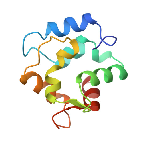
Metal-ion affinity and specificity in EF-hand proteins: coordination geometry and domain plasticity in parvalbumin.
Cates, M.S., Berry, M.B., Ho, E.L., Li, Q., Potter, J.D., Phillips Jr., G.N.(1999) Structure 7: 1269-1278
- PubMed: 10545326 Search on PubMed
- DOI: https://doi.org/10.1016/s0969-2126(00)80060-x
- Primary Citation Related Structures:
1B8C, 1B8L, 1B8R, 1B9A - PubMed Abstract:
The EF-hand family is a large set of Ca(2+)-binding proteins that contain characteristic helix-loop-helix binding motifs that are highly conserved in sequence. Members of this family include parvalbumin and many prominent regulatory proteins such as calmodulin and troponin C. EF-hand proteins are involved in a variety of physiological processes including cell-cycle regulation, second messenger production, muscle contraction, microtubule organization and vision. We have determined the structures of parvalbumin mutants designed to explore the role of the last coordinating residue of the Ca(2+)-binding loop. An E101D substitution has been made in the parvalbumin EF site. The substitution decreases the Ca(2+)-binding affinity 100-fold and increases the Mg(2+)-binding affinity 10-fold. Both the Ca(2+)- and Mg(2+)-bound structures have been determined, and a structural basis has been proposed for the metal-ion-binding properties. The E101D mutation does not affect the Mg(2+) coordination geometry of the binding loop, but it does pull the F helix 1.1 A towards the loop. The E101D-Ca(2+) structure reveals that this mutant cannot obtain the sevenfold coordination preferred by Ca(2+), presumably because of strain limits imposed by tertiary structure. Analysis of these results relative to previously reported structural information supports a model wherein the characteristics of the last coordinating residue and the plasticity of the Ca(2+)-binding loop delimit the allowable geometries for the coordinating sphere.
- Department of Biochemistry and Cell Biology WM Keck Center for Computational Biology, Rice University, 6100 S. Main Street, Houston, TX 77005, USA.
Organizational Affiliation: