Crystal structure of the human p58 killer cell inhibitory receptor (KIR2DL3) specific for HLA-Cw3-related MHC class I.
Maenaka, K., Juji, T., Stuart, D.I., Jones, E.Y.(1999) Structure 7: 391-398
- PubMed: 10196125 Search on PubMed
- DOI: https://doi.org/10.1016/s0969-2126(99)80052-5
- Primary Citation Related Structures:
1B6U - PubMed Abstract:
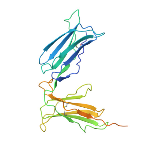
T cells and natural killer (NK) cells perform complementary roles in the cellular immune system. T cells identify infected cells directly through recognition of antigenic peptides that are displayed at the target cell surface by the classical major histocompatibility complex (MHC) class I molecules. NK cells monitor the target cell surface for malfunction of this display system, lysing potentially infected cells that might otherwise evade recognition by the T cells. Human killer cell inhibitory receptors (KIRs) control this process by either inhibiting or activating the cytotoxic activity of NK cells via specific binding to MHC class I molecules on the target cell. We report the crystal structure of the extracellular region of the human p58 KIR (KIR2DL3), which is specific for the human MHC class I molecule HLA-Cw3 and related alleles. The structure shows the predicted topology of two tandem immunoglobulin-like domains, but comparison with the previously reported structure of the related receptor KIR2DL1 reveals an unexpected change of 23 degrees in the relative orientation of these domains. The altered orientation of the immunoglobulin-like domains maintains an unusually acute interdomain elbow angle, which therefore appears to be a distinctive feature of the KIRs. The putative MHC class I binding site is located on the outer surface of the elbow, spanning both domains. The unexpected observation that this binding site can be modulated by differences in the relative domain orientations has implications for the general mechanism of KIR-MHC class I complex formation.
- Laboratory of Molecular Biophysics, Rex Richards Building, South Parks Road, Oxford OX1 3QU, UK. katsumi@biop.ox.ac.uk
Organizational Affiliation: