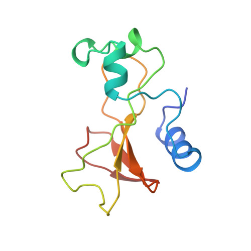
1B2Z

DELETION OF A BURIED SALT BRIDGE IN BARNASE


Experimental Data Snapshot

  • Method: X-RAY DIFFRACTION
  • Resolution: 2.03 Å
  • R-Value Free: 0.228 
  • R-Value Work: 0.170 

wwPDB Validation   3D Report Full Report


This is version 1.5 of the entry. See complete history


Literature

A structural double-mutant cycle: estimating the strength of a buried salt bridge in barnase.

Vaughan, C.K.Harryson, P.Buckle, A.M.Fersht, A.R.

(2002) Acta Crystallogr D Biol Crystallogr 58: 591-600

  • DOI: https://doi.org/10.1107/s0907444902001567
  • Primary Citation of Related Structures:  
    1B20, 1B21, 1B2X, 1B2Z

  • PubMed Abstract: 

    Double-mutant cycles are widely used in the field of protein engineering to measure intermolecular and intramolecular interactions. Ideally, there should be no structural rearrangement of the protein on making the two single mutations and the double mutation within the cycle. However, structural pertubation on mutation does not preclude the use of this method, providing the sum of the changes in the single mutants equals the change in the double mutant. In this way, the energy associated with any structural rearrangement cancels in the double-mutant cycle. Previously, the contribution of a buried salt bridge between Arg69 and Asp93 in barnase to the stability of the folded protein has been determined by double-mutant cycle analysis. In order to determine whether the measured interaction of -14.0 kJ mol(-1) represents the true interaction energy, the crystal structure of each mutant within the double-mutant cycle was solved. Although mutation results in structural shifts, the majority of those in the single mutants are also found in the double mutant; their energetic effects in the double-mutant cycle are therefore cancelled. This study highlights the robust nature of the double-mutant cycle analysis.


  • Organizational Affiliation

    Section of Structural Biology, The Institute of Cancer Research, Chester Beatty Laboratories, 237 Fulham Road, London SW3 6JB, England.


Macromolecules
Find similar proteins by:  (by identity cutoff)  |  3D Structure
Entity ID: 1
MoleculeChains Sequence LengthOrganismDetailsImage
PROTEIN (BARNASE)
A, B, C
110Bacillus amyloliquefaciensMutation(s): 1 
EC: 3.1.27.3
UniProt
Find proteins for P00648 (Bacillus amyloliquefaciens)
Explore P00648 
Go to UniProtKB:  P00648
Entity Groups  
Sequence Clusters30% Identity50% Identity70% Identity90% Identity95% Identity100% Identity
UniProt GroupP00648
Sequence Annotations
Expand
  • Reference Sequence
Small Molecules
Ligands 1 Unique
IDChains Name / Formula / InChI Key2D Diagram3D Interactions
ZN
Query on ZN

Download Ideal Coordinates CCD File 
D [auth C]ZINC ION
Zn
PTFCDOFLOPIGGS-UHFFFAOYSA-N
Experimental Data & Validation

Experimental Data

  • Method: X-RAY DIFFRACTION
  • Resolution: 2.03 Å
  • R-Value Free: 0.228 
  • R-Value Work: 0.170 
  • Space Group: P 32
Unit Cell:
Length ( Å )Angle ( ˚ )
a = 57.75α = 90
b = 57.75β = 90
c = 81.28γ = 120
Software Package:
Software NamePurpose
X-PLORmodel building
REFMACrefinement
iMOSFLMdata reduction
CCP4data scaling
X-PLORphasing

Structure Validation

View Full Validation Report


Entry History 

Deposition Data

Revision History  (Full details and data files)

  • Version 1.0: 1998-12-09
    Type: Initial release
  • Version 1.1: 2008-04-26
    Changes: Version format compliance
  • Version 1.2: 2011-07-13
    Changes: Version format compliance
  • Version 1.3: 2017-10-04
    Changes: Advisory, Refinement description
  • Version 1.4: 2021-11-03
    Changes: Advisory, Database references, Derived calculations
  • Version 1.5: 2023-12-27
    Changes: Data collection