1B2S

STRUCTURAL RESPONSE TO MUTATION AT A PROTEIN-PROTEIN INTERFACE


Experimental Data Snapshot

  • Method: X-RAY DIFFRACTION
  • Resolution: 1.82 Å
  • R-Value Free: 0.249 
  • R-Value Work: 0.194 

wwPDB Validation   3D Report Full Report


This is version 1.3 of the entry. See complete history


Literature

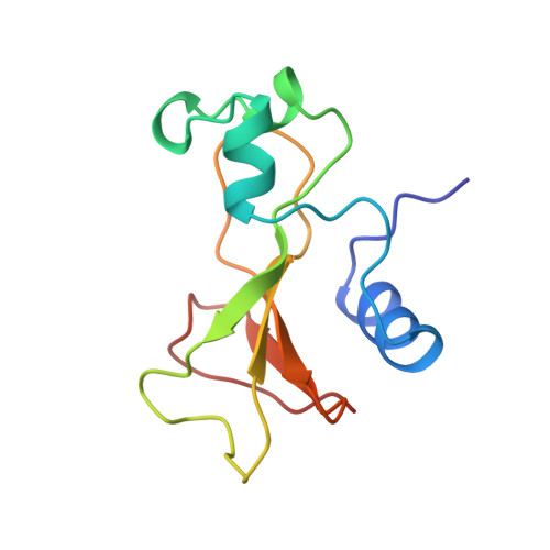
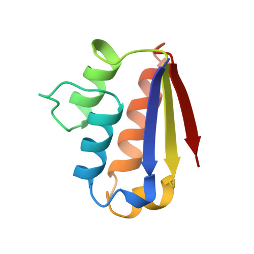
Structural response to mutation at a protein-protein interface.

Vaughan, C.K.Buckle, A.M.Fersht, A.R.

(1999) J Mol Biol 286: 1487-1506

  • DOI: 10.1006/jmbi.1998.2559
  • Primary Citation of Related Structures:  
    1B27, 1B2S, 1B2U, 1B3S

  • PubMed Abstract: 
  • We have crystallised three mutants of the barnase-barstar complex in which interactions across the interface have been deleted by simultaneous mutation of both residues involved in the interaction. Each mutant deletes a different type of interaction at the interface: the first complex bnHis102-->Ala-bsTyr29-->Phe (bn, barnase; bs, barstar), deletes a van der Waals packing interaction; the second complex, bnLys27-->Ala-bsThr42-->Ala, deletes a hydrogen bond; the third, bnLys27-->Ala-bsAsp35-->Ala, deletes a long-range charge-charge interaction ...

    We have crystallised three mutants of the barnase-barstar complex in which interactions across the interface have been deleted by simultaneous mutation of both residues involved in the interaction. Each mutant deletes a different type of interaction at the interface: the first complex bnHis102-->Ala-bsTyr29-->Phe (bn, barnase; bs, barstar), deletes a van der Waals packing interaction; the second complex, bnLys27-->Ala-bsThr42-->Ala, deletes a hydrogen bond; the third, bnLys27-->Ala-bsAsp35-->Ala, deletes a long-range charge-charge interaction. The contribution of each of these side-chains to the stability of the complex is known; the coupling energy between the deleted side-chains is also known. Despite each of the double mutants being significantly destabilised compared with the wild-type, the effects of mutation are local. Only small movements in the main-chain surrounding the sites of mutation and some larger movements of neighbouring side-chains are observed in the mutant complexes. The exact response to mutation is context-dependent and for the same mutant can vary depending upon the environment within the crystal. In some double mutant complexes, interfacial pockets, which are accessible to bulk solvent are formed, whereas interfacial cavities which are isolated from bulk solvent, are formed in others. In all double mutants, water molecules fill the created pockets and cavities. These water molecules mimic the deleted side-chains by occupying positions close to the non-carbon atoms of truncated side-chains and re-making many hydrogen bonds made by the truncated side-chains in the wild-type. It remains extremely difficult, however, to correlate energetic and structural responses to mutation because of unknown changes in entropy and entropy-enthalpy compensation.


    Related Citations: 
    • Protein-Protein Recognition: Crystal Structural Analysis of a Barnase-Barstar Complex at 2.0-A Resolution
      Buckle, A.M., Schreiber, G., Fersht, A.R.
      (1994) Biochemistry 33: 8878
    • Stability and Function: Two Constraints in the Evolution of Barstar and Other Proteins
      Schreiber, G., Buckle, A.M., Fersht, A.R.
      (1994) Structure 2: 945
    • Recognition between a Bacterial Ribonuclease, Barnase, and its Natural Inhibitor, Barstar
      Guillet, V., Lapthorn, A., Hartley, R.W., Mauguen, Y.
      (1993) Structure 1: 165
    • Interaction of Barnase with its Polypeptide Inhibitor Barstar Studied by Protein Engineering
      Schreiber, G., Fersht, A.R.
      (1993) Biochemistry 32: 5145
    • Molecular Structures of a New Family of Ribonucleases
      Mauguen, Y., Hartley, R.W., Dodson, E.J., Dodson, G.G., Bricogne, G., Chothia, C., Jack, A.
      (1982) Nature 297: 162

    Organizational Affiliation

    MRC Centre for Protein Engineering, Hills Road, Cambridge, CB2 2QH, UK.


Macromolecules
Find similar proteins by:  (by identity cutoff)  |  3D Structure
Entity ID: 1
MoleculeChainsSequence LengthOrganismDetailsImage
PROTEIN (BARNASE)A, B, C110Bacillus amyloliquefaciensMutation(s): 1 
EC: 3.1.27.3 (PDB Primary Data), 3.1.27 (UniProt)
UniProt
Find proteins for P00648 (Bacillus amyloliquefaciens)
Explore P00648 
Go to UniProtKB:  P00648
Entity Groups  
Sequence Clusters30% Identity50% Identity70% Identity90% Identity95% Identity100% Identity
UniProt GroupP00648
Protein Feature View
Expand
  • Reference Sequence
Find similar proteins by:  (by identity cutoff)  |  3D Structure
Entity ID: 2
MoleculeChainsSequence LengthOrganismDetailsImage
PROTEIN (BARSTAR)D, E, F90Bacillus amyloliquefaciensMutation(s): 1 
UniProt
Find proteins for P11540 (Bacillus amyloliquefaciens)
Explore P11540 
Go to UniProtKB:  P11540
Entity Groups  
Sequence Clusters30% Identity50% Identity70% Identity90% Identity95% Identity100% Identity
UniProt GroupP11540
Protein Feature View
Expand
  • Reference Sequence
Small Molecules
Ligands 1 Unique
IDChainsName / Formula / InChI Key2D Diagram3D Interactions
SO4
Query on SO4

Download Ideal Coordinates CCD File 
G [auth B],
H [auth E]
SULFATE ION
O4 S
QAOWNCQODCNURD-UHFFFAOYSA-L
 Ligand Interaction
Experimental Data & Validation

Experimental Data

  • Method: X-RAY DIFFRACTION
  • Resolution: 1.82 Å
  • R-Value Free: 0.249 
  • R-Value Work: 0.194 
  • Space Group: C 1 2 1
Unit Cell:
Length ( Å )Angle ( ˚ )
a = 206.24α = 90
b = 43.51β = 107.42
c = 83.69γ = 90
Software Package:
Software NamePurpose
AMoREphasing
REFMACrefinement
MOSFLMdata reduction
CCP4data scaling

Structure Validation

View Full Validation Report


Entry History 

Deposition Data

Revision History  (Full details and data files)

  • Version 1.0: 1998-12-08
    Type: Initial release
  • Version 1.1: 2008-04-26
    Changes: Version format compliance
  • Version 1.2: 2011-07-13
    Changes: Version format compliance
  • Version 1.3: 2021-11-03
    Changes: Data collection, Database references, Derived calculations