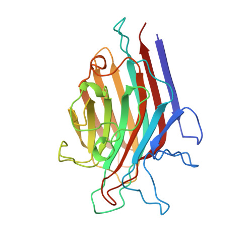
1AXZ

ERYTHRINA CORALLODENDRON LECTIN IN COMPLEX WITH D-GALACTOSE


Experimental Data Snapshot

  • Method: X-RAY DIFFRACTION
  • Resolution: 1.95 Å
  • R-Value Free: 0.206 
  • R-Value Work: 0.179 
  • R-Value Observed: 0.179 

Starting Model: experimental
View more details

wwPDB Validation   3D Report Full Report


Ligand Structure Quality Assessment 


This is version 2.1 of the entry. See complete history


Literature

Structures of the Erythrina corallodendron lectin and of its complexes with mono- and disaccharides.

Elgavish, S.Shaanan, B.

(1998) J Mol Biol 277: 917-932

  • DOI: https://doi.org/10.1006/jmbi.1998.1664
  • Primary Citation of Related Structures:  
    1AX0, 1AX1, 1AX2, 1AXY, 1AXZ

  • PubMed Abstract: 

    The structures of the Erythrina corallodendron lectin (EcorL) and of its complexes with galactose, N-acetylgalactosamine, lactose and N-acetyllactosamine were determined at a resolution of 1.9 to 1.95 A. The final R-values of the five models are in the range 0.169 to 0.181. The unusual, non-canonical, dimer interface of EcorL is made of beta-strands from the two monomers, which face one another in a "hand-shake" mode. The galactose molecule in the primary binding site is bound in an identical way in all four complexes. Features of the electrostatic potential of the galactose molecule match those of the potential in the combining site, thus probably pointing to the contribution of the electrostatic energy to determining the orientation of the ligand. No conformational change occurs in the protein upon binding the ligand. Subtle variations in the binding mode of the second monosaccharide (glucose in the complex with lactose and N-acetylglucosamine in the complex with N-acetyllactosamine) were observed. The mobility of Gln219 is lower in the complexes with the disaccharides than in the complexes with the monosaccharides, indicating further recruitment of this residue to ligand binding through more extensive hydrogen bonding in the former complexes. Water molecules that have been located in the combining sites of the five structures undergo rearrangement in response to binding of the different ligands. The new structural information is in qualitative agreement with thermodynamic data on the binding to EcorL.


  • Organizational Affiliation

    The Institute of Life Sciences, The Hebrew University of Jerusalem, Givat Ram, Israel.


Macromolecules
Find similar proteins by:  (by identity cutoff)  |  3D Structure
Entity ID: 1
MoleculeChains Sequence LengthOrganismDetailsImage
LECTIN239Erythrina corallodendronMutation(s): 0 
UniProt
Find proteins for P16404 (Erythrina corallodendron)
Explore P16404 
Go to UniProtKB:  P16404
Entity Groups  
Sequence Clusters30% Identity50% Identity70% Identity90% Identity95% Identity100% Identity
UniProt GroupP16404
Glycosylation
Glycosylation Sites: 1
Sequence Annotations
Expand
  • Reference Sequence
Oligosaccharides

Help

Entity ID: 2
MoleculeChains Length2D Diagram Glycosylation3D Interactions
beta-D-xylopyranose-(1-2)-[alpha-D-mannopyranose-(1-3)][alpha-D-mannopyranose-(1-6)]beta-D-mannopyranose-(1-4)-2-acetamido-2-deoxy-beta-D-glucopyranose-(1-4)-[alpha-L-fucopyranose-(1-3)]2-acetamido-2-deoxy-beta-D-glucopyranose
B
7N-Glycosylation
Glycosylation Resources
GlyTouCan:  G26295XE
GlyCosmos:  G26295XE
GlyGen:  G26295XE
Small Molecules
Binding Affinity Annotations 
IDSourceBinding Affinity
GAL PDBBind:  1AXZ Kd: 6.37e+5 (nM) from 1 assay(s)
Experimental Data & Validation

Experimental Data

  • Method: X-RAY DIFFRACTION
  • Resolution: 1.95 Å
  • R-Value Free: 0.206 
  • R-Value Work: 0.179 
  • R-Value Observed: 0.179 
  • Space Group: C 1 2 1
Unit Cell:
Length ( Å )Angle ( ˚ )
a = 84.202α = 90
b = 72.99β = 113.44
c = 71.29γ = 90
Software Package:
Software NamePurpose
X-PLORmodel building
CNSrefinement
X-PLORrefinement
DENZOdata reduction
SCALEPACKdata scaling
X-PLORphasing

Structure Validation

View Full Validation Report


Ligand Structure Quality Assessment 


Entry History 

Deposition Data

Revision History  (Full details and data files)

  • Version 1.0: 1998-05-06
    Type: Initial release
  • Version 1.1: 2008-03-10
    Changes: Version format compliance
  • Version 1.2: 2011-07-13
    Changes: Non-polymer description, Version format compliance
  • Version 1.3: 2011-11-16
    Changes: Atomic model
  • Version 2.0: 2020-07-29
    Type: Remediation
    Reason: Carbohydrate remediation
    Changes: Atomic model, Data collection, Derived calculations, Other, Structure summary
  • Version 2.1: 2024-04-03
    Changes: Data collection, Database references, Refinement description, Structure summary