The solution structure of oxidized rat microsomal cytochrome b5.
Arnesano, F., Banci, L., Bertini, I., Felli, I.C.(1998) Biochemistry 37: 173-184
- PubMed: 9425037 Search on PubMed
- DOI: https://doi.org/10.1021/bi971896w
- Primary Citation Related Structures:
1AW3, 1AXX, 2AXX - PubMed Abstract:
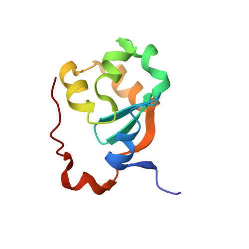
The solution structure of oxidized rat microsomal cytochrome b5 has been obtained from 1H NMR spectra measured at 800 MHz. The available assignment has been extended to 78% of the total protons and 95% of the residues. From 1372 meaningful NOEs, a family of 40 structures has been obtained through the program DYANA; 235 pseudocontact shifts have been then added as further constraints, obtaining an essentially similar family of structures. This latter family has been further refined through restrained energy minimization. The final RMSD values with respect to the average structure are 0.58 +/- 0.10 A and 1.05 +/- 0.11 A for backbone and heavy atoms, respectively. The high quality of the structure allows meaningful comparisons with the solution structure of the reduced protein, with the X-ray and solution structures of the oxidized bovine isoenzyme, and with the solution structure of the apoprotein. Upon loss of one electron, the heme plane undergoes a change in its orientation, possibly due to the change of the total charge. Propionate 7 appears to have a conformation which is dependent on the oxidation state of the iron. Helices alpha2 and alpha4 also experience changes in their average positions in the two oxidation states. Finally, the backbone NHs experience different exchange properties in the two oxidation states. While those present in the beta sheets forming the basis of the heme pocket are nonexchanging in both oxidation states, the NHs in the helices forming the heme-binding pocket are exchanging with the bulk solvent in the oxidized form, indicating larger local mobility in this state. This observation could suggest that, to optimize the electron transfer process, the local mobility should be properly tuned.
- Department of Chemistry, University of Florence, Italy.
Organizational Affiliation: