1AJ0

CRYSTAL STRUCTURE OF A TERNARY COMPLEX OF E. COLI DIHYDROPTEROATE SYNTHASE


Experimental Data Snapshot

  • Method: X-RAY DIFFRACTION
  • Resolution: 2.00 Å
  • R-Value Work: 0.206 

wwPDB Validation   3D Report Full Report


This is version 1.3 of the entry. See complete history


Literature

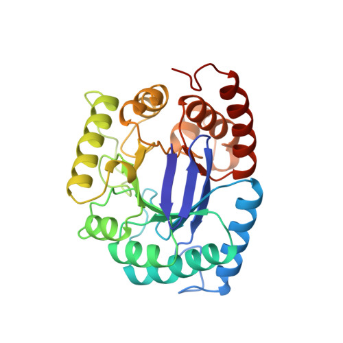
Crystal structure of the anti-bacterial sulfonamide drug target dihydropteroate synthase.

Achari, A.Somers, D.O.Champness, J.N.Bryant, P.K.Rosemond, J.Stammers, D.K.

(1997) Nat Struct Biol 4: 490-497

  • DOI: https://doi.org/10.1038/nsb0697-490
  • Primary Citation of Related Structures:  
    1AJ0, 1AJ2, 1AJZ

  • PubMed Abstract: 

    Sulfonamides were amongst the first clinically useful antibacterial agents to be discovered. The identification of sulfanilamide as the active component of the dye Prontosil rubrum led to the synthesis of clinically useful analogues. Today sulfamethoxazole (in combination with trimethoprim), is used to treat urinary tract infections caused by bacteria such as Escherichia coli and is also a first-line treatment for pneumonia caused by the fungus Pneumocystis carinii, a common condition in AIDS patients. The site of action is the de novo folate biosynthesis enzyme dihydropteroate synthase (DHPS) where sulfonamides act as analogues of one of the substrates, para-aminobenzoic acid (pABA). We report here the crystal structure of E.coli DHPS at 2.0 A resolution refined to an R-factor of 0.185. The single domain of 282 residues forms an eight-stranded alpha/beta-barrel. The 7,8-dihydropterin pyrophosphate (DHPPP) substrate binds in a deep cleft in the barrel, whilst sulfanilamide binds closer to the surface. The DHPPP ligand site is highly conserved amongst prokaryotic and eukaryotic DHPSs.


  • Organizational Affiliation

    Glaxo Wellcome Research & Development, Beckenham, Kent, UK. aa37795@ggr.co.uk


Macromolecules
Find similar proteins by:  (by identity cutoff)  |  3D Structure
Entity ID: 1
MoleculeChains Sequence LengthOrganismDetailsImage
DIHYDROPTEROATE SYNTHASE282Escherichia coliMutation(s): 0 
EC: 2.5.1.15
UniProt
Find proteins for P0AC13 (Escherichia coli (strain K12))
Explore P0AC13 
Go to UniProtKB:  P0AC13
Entity Groups  
Sequence Clusters30% Identity50% Identity70% Identity90% Identity95% Identity100% Identity
UniProt GroupP0AC13
Sequence Annotations
Expand
  • Reference Sequence
Small Molecules
Ligands 3 Unique
IDChains Name / Formula / InChI Key2D Diagram3D Interactions
PH2
Query on PH2

Download Ideal Coordinates CCD File 
C [auth A]2-AMINO-6-HYDROXYMETHYL-7,8-DIHYDRO-3H-PTERIDIN-4-ONE
C7 H9 N5 O2
CQQNNQTXUGLUEV-UHFFFAOYSA-N
SAN
Query on SAN

Download Ideal Coordinates CCD File 
D [auth A]SULFANILAMIDE
C6 H8 N2 O2 S
FDDDEECHVMSUSB-UHFFFAOYSA-N
SO4
Query on SO4

Download Ideal Coordinates CCD File 
B [auth A]SULFATE ION
O4 S
QAOWNCQODCNURD-UHFFFAOYSA-L
Experimental Data & Validation

Experimental Data

  • Method: X-RAY DIFFRACTION
  • Resolution: 2.00 Å
  • R-Value Work: 0.206 
  • Space Group: C 1 21 1
Unit Cell:
Length ( Å )Angle ( ˚ )
a = 92.91α = 90
b = 60.67β = 114.67
c = 59.82γ = 90
Software Package:
Software NamePurpose
PROFFTrefinement
XENGENdata reduction
XENGENdata scaling

Structure Validation

View Full Validation Report


Entry History 

Deposition Data

Revision History  (Full details and data files)

  • Version 1.0: 1998-05-20
    Type: Initial release
  • Version 1.1: 2008-03-24
    Changes: Version format compliance
  • Version 1.2: 2011-07-13
    Changes: Derived calculations, Version format compliance
  • Version 1.3: 2024-02-07
    Changes: Data collection, Database references, Derived calculations