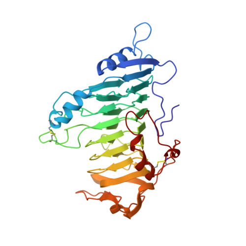
1AIR

PECTATE LYASE C FROM ERWINIA CHRYSANTHEMI (EC16) TO A RESOLUTION OF 2.2 ANGSTROMS WITH 128 WATERS


Experimental Data Snapshot

  • Method: X-RAY DIFFRACTION
  • Resolution: 2.20 Å
  • R-Value Free: 0.223 
  • R-Value Work: 0.172 
  • R-Value Observed: 0.172 

wwPDB Validation   3D Report Full Report


This is version 1.3 of the entry. See complete history


Literature

The Refined Three-Dimensional Structure of Pectate Lyase E from Erwinia chrysanthemi at 2.2 A Resolution.

Lietzke, S.E.Scavetta, R.D.Yoder, M.D.Jurnak, F.

(1996) Plant Physiol 111: 73-92

  • DOI: https://doi.org/10.1104/pp.111.1.73
  • Primary Citation of Related Structures:  
    1AIR

  • PubMed Abstract: 

    The crystal structure of pectate lyase E (PelE; EC 4.2.2.2) from the enterobacteria Erwinia chrysanthemi has been refined by molecular dynamics techniques to a resolution of 2.2 A and an R factor (an agreement factor between observed structure factor amplitudes) of 16.1%. The final model consists of all 355 amino acids and 157 water molecules. The root-mean-square deviation from ideality is 0.009 A for bond lengths and 1.721[deg] for bond angles. The structure of PelE bound to a lanthanum ion, which inhibits the enzymatic activity, has also been refined and compared to the metal-free protein. In addition, the structures of pectate lyase C (PelC) in the presence and absence of a lutetium ion have been refined further using an improved algorithm for identifying waters and other solvent molecules. The two putative active site regions of PelE have been compared to those in the refined structure of PelC. The analysis of the atomic details of PelE and PelC in the presence and absence of lanthanide ions provides insight into the enzymatic mechanism of pectate lyases.


  • Organizational Affiliation

    Department of Biochemistry, University of California, Riverside, California 92521.


Macromolecules
Find similar proteins by:  (by identity cutoff)  |  3D Structure
Entity ID: 1
MoleculeChains Sequence LengthOrganismDetailsImage
PECTATE LYASE C353Dickeya chrysanthemiMutation(s): 0 
EC: 4.2.2.2
UniProt
Find proteins for P11073 (Dickeya chrysanthemi)
Explore P11073 
Go to UniProtKB:  P11073
Entity Groups  
Sequence Clusters30% Identity50% Identity70% Identity90% Identity95% Identity100% Identity
UniProt GroupP11073
Sequence Annotations
Expand
  • Reference Sequence
Small Molecules
Ligands 1 Unique
IDChains Name / Formula / InChI Key2D Diagram3D Interactions
SO4
Query on SO4

Download Ideal Coordinates CCD File 
B [auth A],
C [auth A]
SULFATE ION
O4 S
QAOWNCQODCNURD-UHFFFAOYSA-L
Experimental Data & Validation

Experimental Data

  • Method: X-RAY DIFFRACTION
  • Resolution: 2.20 Å
  • R-Value Free: 0.223 
  • R-Value Work: 0.172 
  • R-Value Observed: 0.172 
  • Space Group: P 21 21 21
Unit Cell:
Length ( Å )Angle ( ˚ )
a = 72α = 90
b = 83β = 90
c = 95γ = 90
Software Package:
Software NamePurpose
SDMSdata collection
SDMSdata reduction
X-PLORmodel building
X-PLORrefinement
SDMSdata scaling
X-PLORphasing

Structure Validation

View Full Validation Report


Entry History 

Deposition Data

Revision History  (Full details and data files)

  • Version 1.0: 1997-06-16
    Type: Initial release
  • Version 1.1: 2008-03-24
    Changes: Version format compliance
  • Version 1.2: 2011-07-13
    Changes: Version format compliance
  • Version 1.3: 2024-06-05
    Changes: Data collection, Database references, Derived calculations, Other