1AGI

CRYSTAL STRUCTURE OF BOVINE ANGIOGENIN AT 1.5 ANGSTROMS RESOLUTION


Experimental Data Snapshot

  • Method: X-RAY DIFFRACTION
  • Resolution: 1.50 Å
  • R-Value Work: 0.203 
  • R-Value Observed: 0.203 

wwPDB Validation   3D Report Full Report


This is version 1.2 of the entry. See complete history


Literature

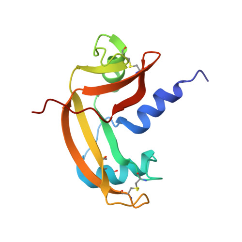
Crystal structure of bovine angiogenin at 1.5-A resolution.

Acharya, K.R.Shapiro, R.Riordan, J.F.Vallee, B.L.

(1995) Proc Natl Acad Sci U S A 92: 2949-2953

  • DOI: https://doi.org/10.1073/pnas.92.7.2949
  • Primary Citation of Related Structures:  
    1AGI

  • PubMed Abstract: 

    The capacity of angiogenin (Ang) to induce blood vessel growth is critically dependent on its ribonucleolytic activity. Crystallography and mutagenesis of human Ang have previously shown that its pyrimidine binding site is obstructed by Gln-117, implying that a conformational change is a key part of the mechanism of Ang action. The 1.5-A-resolution crystal structure of bovine Ang, in which glutamic acid is substituted for Gln-117, now confirms that a blocked active site is characteristic of these proteins. Indeed, the inactive conformation of bovine Ang is stabilized by a more extensive set of interactions than is that of human Ang. The three-dimensional structure of the putative receptor binding site is also well conserved in the two proteins. The Arg-Gly-Asp segment of this site in bovine Ang, which is replaced by Arg-Glu-Asn in human Ang, does not have a conformation typical of an integrin recognition site.


  • Organizational Affiliation

    School of Biology and Biochemistry, University of Bath, United Kingdom.


Macromolecules
Find similar proteins by:  (by identity cutoff)  |  3D Structure
Entity ID: 1
MoleculeChains Sequence LengthOrganismDetailsImage
ANGIOGENIN125Bos taurusMutation(s): 0 
EC: 3.1.27
UniProt
Find proteins for P10152 (Bos taurus)
Explore P10152 
Go to UniProtKB:  P10152
Entity Groups  
Sequence Clusters30% Identity50% Identity70% Identity90% Identity95% Identity100% Identity
UniProt GroupP10152
Sequence Annotations
Expand
  • Reference Sequence
Experimental Data & Validation

Experimental Data

  • Method: X-RAY DIFFRACTION
  • Resolution: 1.50 Å
  • R-Value Work: 0.203 
  • R-Value Observed: 0.203 
  • Space Group: P 21 21 21
Unit Cell:
Length ( Å )Angle ( ˚ )
a = 30.69α = 90
b = 54.45β = 90
c = 74.75γ = 90
Software Package:
Software NamePurpose
X-PLORmodel building
X-PLORrefinement
X-PLORphasing

Structure Validation

View Full Validation Report


Entry History 

Deposition Data

Revision History  (Full details and data files)

  • Version 1.0: 1996-04-03
    Type: Initial release
  • Version 1.1: 2008-03-03
    Changes: Version format compliance
  • Version 1.2: 2011-07-13
    Changes: Version format compliance