1AEI | pdb_00001aei

CRYSTAL STRUCTURE OF THE ANNEXIN XII HEXAMER


Experimental Data Snapshot

  • Method: X-RAY DIFFRACTION
  • Resolution: 2.80 Å
  • R-Value Free: 
    0.278 (Depositor) 
  • R-Value Work: 
    0.236 (Depositor) 
  • R-Value Observed: 
    0.236 (Depositor) 

Starting Model: experimental
View more details

wwPDB Validation 3D Report Full Report

Validation slider image for 1AEI

This is version 1.4 of the entry. See complete history

Literature

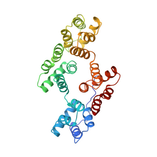
Crystal structure of the annexin XII hexamer and implications for bilayer insertion.

Luecke, H.Chang, B.T.Mailliard, W.S.Schlaepfer, D.D.Haigler, H.T.

(1995) Nature 378: 512-515

  • DOI: https://doi.org/10.1038/378512a0
  • Primary Citation Related Structures: 
    1AEI

  • PubMed Abstract: 

    Annexins are a family of calcium- and phospholipid-binding proteins implicated in a number of biological processes including membrane fusion and ion channel formation. The crystal structure of the annexin XII hexamer, refined at 2.8 A resolution, forms a concave disk with 3-2 symmetry, about 100 A in diameter and 70 A thick with a central hydrophilic pore. Six intermolecular Ca2+ ions are involved in hexamer formation. An additional 18 Ca2+ ions are located on the perimeter of the disk, accessible only from the side of the hexameric disk. On the basis of the hexamer structure we propose here a new mode of protein-phospholipid bilayer interaction that is distinct from the hydrophobic insertion of typical membrane proteins. This speculative model postulates the Ca(2+)-dependent insertion of the hydrophilic annexin XII hexamer into phospholipid bilayers with local reorientation of the bilayer phospholipids.


  • Organizational Affiliation
    • Stanford Synchrotron Radiation Laboratory, California 94305, USA.

Macromolecule Content 

  • Total Structure Weight: 211.11 kDa 
  • Atom Count: 14,796 
  • Modeled Residue Count: 1,890 
  • Deposited Residue Count: 1,890 
  • Unique protein chains: 1

Macromolecules

Find similar proteins by:|  3D Structure
Entity ID: 1
MoleculeChains  Sequence LengthOrganismDetailsImage
ANNEXIN XII
A, B, C, D, E
A, B, C, D, E, F
315Hydra vulgarisMutation(s): 0 
UniProt
Find proteins for P26256 (Hydra vulgaris)
Explore P26256 
Go to UniProtKB:  P26256
Entity Groups
Sequence Clusters30% Identity50% Identity70% Identity90% Identity95% Identity100% Identity
UniProt GroupP26256
Sequence Annotations
Expand
Reference Sequence

Small Molecules

Ligands 1 Unique
IDChains Name / Formula / InChI Key2D Diagram3D Interactions
CA

Query on CA


Download:Ideal Coordinates CCD File
AA [auth F]
BA [auth F]
CA [auth F]
DA [auth F]
G [auth A]
AA [auth F],
BA [auth F],
CA [auth F],
DA [auth F],
G [auth A],
H [auth A],
I [auth A],
J [auth A],
K [auth B],
L [auth B],
M [auth B],
N [auth B],
O [auth C],
P [auth C],
Q [auth C],
R [auth C],
S [auth D],
T [auth D],
U [auth D],
V [auth D],
W [auth E],
X [auth E],
Y [auth E],
Z [auth E]
CALCIUM ION
Ca
BHPQYMZQTOCNFJ-UHFFFAOYSA-N

Experimental Data & Validation

Experimental Data

  • Method: X-RAY DIFFRACTION
  • Resolution: 2.80 Å
  • R-Value Free:  0.278 (Depositor) 
  • R-Value Work:  0.236 (Depositor) 
  • R-Value Observed: 0.236 (Depositor) 
Space Group: P 1 21 1
Unit Cell:
Length ( Å )Angle ( ˚ )
a = 71.2α = 90
b = 179.2β = 97.6
c = 100.7γ = 90
Software Package:
Software NamePurpose
GLRFphasing
X-PLORmodel building
X-PLORrefinement
MOSFLMdata reduction
CCP4data scaling
ROTAVATAdata scaling
X-PLORphasing

Structure Validation

View Full Validation Report


Entry History 

Deposition Data

Revision History  (Full details and data files)

  • Version 1.0: 1997-08-20
    Type: Initial release
  • Version 1.1: 2008-03-24
    Changes: Version format compliance
  • Version 1.2: 2011-07-13
    Changes: Version format compliance
  • Version 1.3: 2024-02-07
    Changes: Data collection, Database references, Derived calculations, Refinement description
  • Version 1.4: 2024-04-03
    Changes: Refinement description