1A7D

CHLOROMET MYOHEMERYTHRIN FROM THEMISTE ZOSTERICOLA


Experimental Data Snapshot

  • Method: X-RAY DIFFRACTION
  • Resolution: 1.80 Å
  • R-Value Free: 0.227 
  • R-Value Work: 0.180 
  • R-Value Observed: 0.180 

wwPDB Validation   3D Report Full Report


This is version 1.3 of the entry. See complete history


Literature

Structures of wild-type chloromet and L103N hydroxomet Themiste zostericola myohemerythrins at 1.8 A resolution.

Martins, L.J.Hill, C.P.Ellis Jr., W.R.

(1997) Biochemistry 36: 7044-7049

  • DOI: https://doi.org/10.1021/bi9630422
  • Primary Citation of Related Structures:  
    1A7D, 1A7E

  • PubMed Abstract: 

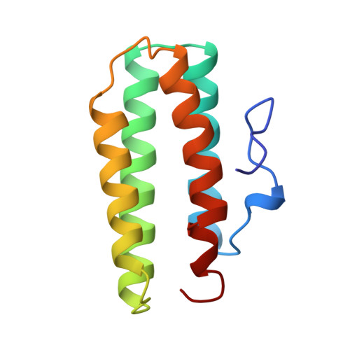
    Myohemerythrin (Mhr) is a nonheme iron oxygen carrier found in the retractor muscles of marine "peanut" worms. The X-ray crystal structures of two recombinant Themiste zostericola Mhrs are reported to a resolution of 1.8 A. Surprisingly, the met wild-type structure (R = 17.8%) was found to contain chloride bound to Fe2, while coordinated hydroxide was found in the met L103N structure (R = 18.3%). An internal water molecule was also found distal to the Fe-O-Fe center of the mutant protein, forming hydrogen bonds with the coordinated hydroxide and the OD1 atom of Asn-103. This finding is consistent with the kinetic and spectroscopic results reported for the L103N mutant Mhr [Raner, G. M., Martins, L. J., & Ellis, W. R., Jr. (1997) Biochemistry 36, 7037-7043]. Possible roles for the side chain of residue 103 (Leu in wild-type Mhr) in gating ligand binding are also discussed.


  • Organizational Affiliation

    Department of Chemistry, University of Utah, Salt Lake City 84112, USA.


Macromolecules
Find similar proteins by:  (by identity cutoff)  |  3D Structure
Entity ID: 1
MoleculeChains Sequence LengthOrganismDetailsImage
MYOHEMERYTHRIN118Themiste hennahiMutation(s): 0 
UniProt
Find proteins for P02247 (Themiste hennahi)
Explore P02247 
Go to UniProtKB:  P02247
Entity Groups  
Sequence Clusters30% Identity50% Identity70% Identity90% Identity95% Identity100% Identity
UniProt GroupP02247
Sequence Annotations
Expand
  • Reference Sequence
Experimental Data & Validation

Experimental Data

  • Method: X-RAY DIFFRACTION
  • Resolution: 1.80 Å
  • R-Value Free: 0.227 
  • R-Value Work: 0.180 
  • R-Value Observed: 0.180 
  • Space Group: P 21 21 21
Unit Cell:
Length ( Å )Angle ( ˚ )
a = 41.842α = 90
b = 81.26β = 90
c = 36.831γ = 90
Software Package:
Software NamePurpose
X-PLORmodel building
X-PLORrefinement
R-AXISdata reduction
R-AXISdata scaling
X-PLORphasing

Structure Validation

View Full Validation Report


Entry History 

Deposition Data

Revision History  (Full details and data files)

  • Version 1.0: 1998-10-14
    Type: Initial release
  • Version 1.1: 2008-03-24
    Changes: Version format compliance
  • Version 1.2: 2011-07-13
    Changes: Version format compliance
  • Version 1.3: 2024-02-07
    Changes: Data collection, Database references, Derived calculations, Other