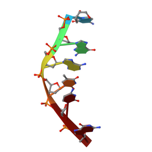
Water ring structure at DNA interfaces: hydration and dynamics of DNA-anthracycline complexes.
Lipscomb, L.A., Peek, M.E., Zhou, F.X., Bertrand, J.A., VanDerveer, D., Williams, L.D.(1994) Biochemistry 33: 3649-3659
- PubMed: 8142363 Search on PubMed
- DOI: https://doi.org/10.1021/bi00178a023
- Primary Citation Related Structures:
151D, 152D - PubMed Abstract:
In crystallographic structures of biological macromolecules, one can observe many hydration rings that originate at one water molecule, pass via hydrogen bonds through several others, and return to the original water molecule. Five-membered water rings have been thought to occur with greater frequency than other ring sizes. We describe a quantitative assessment of relationships between water ring size and frequency of occurrence in the vicinity of nucleic acid interfaces. This report focuses on low-temperature X-ray crystallographic structures of two anthracyclines, adriamycin (ADRI) and daunomycin (DAUN), bound to d(CGATCG) and on several DNA structures published previously by others. We have obtained excellent low-temperature (-160 degrees C, LT) X-ray intensity data for d(CGATCG)-adriamycin and d(CGATCG)-daunomycin with a multiwire area detector. The LTX-ray data sets contain 20% (daunomycin, LT-DAUN) and 35% (adriamycin, LT-ADRI) more reflections than were used to derive the original room-temperature (15 degrees C) structures [Frederick, C.A., Williams, L.D., Ughetto, G., van der Marel, G. A., van Boom, J.H., Rich, A., & Wang, A.H.-J. (1990) Biochemistry 29, 2538-2549]. The results show that five-membered water rings are not preferred over other ring sizes. This assessment is consistent with our observation of broad dispersion W-W-W angles (sigma = 20 degrees). In addition, we report that the thermal mobility, distinct from the static disorder, of the amino sugar of daunomycin and adriamycin is significantly greater than that of the rest of the complex. This mobility implies that if the central AT base pair is switched to a CG base pair, there should be a low energy cost in avoiding the guanine amino group. The energy difference (for the sugar-binding preference) between d(CGTACG) and d(CGCGCG) could be considerably less than 20 kcal/mol, a value proposed previously from computation.
- School of Chemistry & Biochemistry, Georgia Institute of Technology, Atlanta 30332.
Organizational Affiliation: