11GS

Glutathione s-transferase complexed with ethacrynic acid-glutathione conjugate (form ii)


Experimental Data Snapshot

  • Method: X-RAY DIFFRACTION
  • Resolution: 2.30 Å
  • R-Value Free: 0.270 
  • R-Value Work: 0.214 
  • R-Value Observed: 0.214 

wwPDB Validation   3D Report Full Report


Ligand Structure Quality Assessment 


This is version 1.2 of the entry. See complete history


Literature

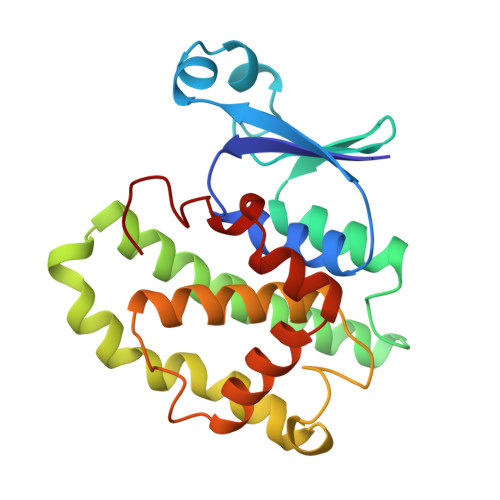
The glutathione conjugate of ethacrynic acid can bind to human pi class glutathione transferase P1-1 in two different modes.

Oakley, A.J.Lo Bello, M.Mazzetti, A.P.Federici, G.Parker, M.W.

(1997) FEBS Lett 419: 32-36

  • DOI: https://doi.org/10.1016/s0014-5793(97)01424-5
  • Primary Citation of Related Structures:  
    11GS

  • PubMed Abstract: 
  • The diuretic drug ethacrynic acid, an inhibitor of pi class glutathione S-transferase, has been tested in clinical trials as an adjuvant in chemotherapy. We recently solved the crystal structure of this enzyme in complex with ethacrynic acid and its glutathione conjugate ...

    The diuretic drug ethacrynic acid, an inhibitor of pi class glutathione S-transferase, has been tested in clinical trials as an adjuvant in chemotherapy. We recently solved the crystal structure of this enzyme in complex with ethacrynic acid and its glutathione conjugate. Here we present a new structure of the ethacrynic-glutathione conjugate complex. In this structure the ethacrynic moiety of the complex is shown to bind in a completely different orientation to that previously observed. Thus there are at least two binding modes possible, an observation of great importance to the design of second generation inhibitors of the enzyme.


    Organizational Affiliation

    The Ian Potter Foundation Protein Crystallography Laboratory, St. Vincent's Institute of Medical Research, Fitzroy, Vic., Australia.


Macromolecules
Find similar proteins by:  (by identity cutoff)  |  3D Structure
Entity ID: 1
MoleculeChainsSequence LengthOrganismDetailsImage
GLUTATHIONE S-TRANSFERASE
A, B
210Homo sapiensMutation(s): 0 
Gene Names: GSTP1FAEES3GST3
EC: 2.5.1.18
UniProt & NIH Common Fund Data Resources
Find proteins for P09211 (Homo sapiens)
Explore P09211 
Go to UniProtKB:  P09211
PHAROS:  P09211
Entity Groups  
Sequence Clusters30% Identity50% Identity70% Identity90% Identity95% Identity100% Identity
UniProt GroupP09211
Protein Feature View
Expand
  • Reference Sequence
Small Molecules
Ligands 3 Unique
IDChainsName / Formula / InChI Key2D Diagram3D Interactions
GSH
Query on GSH

Download Ideal Coordinates CCD File 
C [auth A],
F [auth B]
GLUTATHIONE
C10 H17 N3 O6 S
RWSXRVCMGQZWBV-WDSKDSINSA-N
 Ligand Interaction
EAA
Query on EAA

Download Ideal Coordinates CCD File 
D [auth A],
G [auth B]
ETHACRYNIC ACID
C13 H12 Cl2 O4
AVOLMBLBETYQHX-UHFFFAOYSA-N
 Ligand Interaction
MES
Query on MES

Download Ideal Coordinates CCD File 
E [auth A],
H [auth B]
2-(N-MORPHOLINO)-ETHANESULFONIC ACID
C6 H13 N O4 S
SXGZJKUKBWWHRA-UHFFFAOYSA-N
 Ligand Interaction
Experimental Data & Validation

Experimental Data

  • Method: X-RAY DIFFRACTION
  • Resolution: 2.30 Å
  • R-Value Free: 0.270 
  • R-Value Work: 0.214 
  • R-Value Observed: 0.214 
  • Space Group: C 1 2 1
Unit Cell:
Length ( Å )Angle ( ˚ )
a = 77.78α = 90
b = 89.56β = 97.62
c = 68.65γ = 90
Software Package:
Software NamePurpose
X-PLORmodel building
X-PLORrefinement
DENZOdata reduction
SCALEPACKdata scaling
X-PLORphasing

Structure Validation

View Full Validation Report


Ligand Structure Quality Assessment 


Entry History 

Deposition Data

Revision History  (Full details and data files)

  • Version 1.0: 1999-01-13
    Type: Initial release
  • Version 1.1: 2008-03-03
    Changes: Version format compliance
  • Version 1.2: 2011-07-13
    Changes: Version format compliance