7KQS

A 1.68-A resolution 3-fluoro-L-tyrosine bound crystal structure of heme-dependent tyrosine hydroxylase


Experimental Data Snapshot

  • Method: X-RAY DIFFRACTION
  • Resolution: 1.68 Å
  • R-Value Free: 0.203 
  • R-Value Work: 0.172 
  • R-Value Observed: 0.173 

wwPDB Validation   3D Report Full Report


Ligand Structure Quality Assessment 


This is version 1.2 of the entry. See complete history


Literature

Molecular Rationale for Partitioning between C-H and C-F Bond Activation in Heme-Dependent Tyrosine Hydroxylase.

Wang, Y.Davis, I.Shin, I.Xu, H.Liu, A.

(2021) J Am Chem Soc 143: 4680-4693

  • DOI: https://doi.org/10.1021/jacs.1c00175
  • Primary Citation of Related Structures:  
    7KQR, 7KQS, 7KQT, 7KQU

  • PubMed Abstract: 

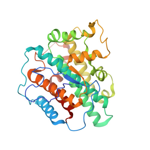
    The heme-dependent l-tyrosine hydroxylases (TyrHs) in natural product biosynthesis constitute a new enzyme family in contrast to the nonheme iron enzymes for DOPA production. A representative TyrH exhibits dual reactivity of C-H and C-F bond cleavage when challenged with 3-fluoro-l-tyrosine (3-F-Tyr) as a substrate. However, little is known about how the enzyme mediates two distinct reactions. Herein, a new TyrH from the thermophilic bacterium Streptomyces sclerotialus (SsTyrH) was functionally and structurally characterized. A de novo crystal structure of the enzyme-substrate complex at 1.89-Å resolution provides the first comprehensive structural study of this hydroxylase. The binding conformation of l-tyrosine indicates that C-H bond hydroxylation is initiated by electron transfer. Mutagenesis studies confirmed that an active site histidine, His88, participates in catalysis. We also obtained a 1.68-Å resolution crystal structure in complex with the monofluorinated substrate, 3-F-Tyr, which shows one binding conformation but two orientations of the fluorine atom with a ratio of 7:3, revealing that the primary factor of product distribution is the substrate orientation. During in crystallo reaction, a ferric-hydroperoxo intermediate (compound 0, Fe 3+ -OOH) was observed with 3-F-Tyr as a substrate based on characteristic spectroscopic features. We determined the crystal structure of this compound 0-type intermediate and refined it to 1.58-Å resolution. Collectively, this study provided the first molecular details of the heme-dependent TyrH and determined the primary factor that dictates the partitioning between the dual reactivities of C-H and C-F bond activation.


  • Organizational Affiliation

    Department of Chemistry, The University of Texas at San Antonio, One UTSA Circle, Texas 78249, United States.


Macromolecules
Find similar proteins by:  (by identity cutoff)  |  3D Structure
Entity ID: 1
MoleculeChains Sequence LengthOrganismDetailsImage
Heme-dependent L-tyrosine hydroxylase
A, B
316Streptomyces sclerotialusMutation(s): 0 
Entity Groups  
Sequence Clusters30% Identity50% Identity70% Identity90% Identity95% Identity100% Identity
Sequence Annotations
Expand
  • Reference Sequence
Small Molecules
Ligands 4 Unique
IDChains Name / Formula / InChI Key2D Diagram3D Interactions
HEM (Subject of Investigation/LOI)
Query on HEM

Download Ideal Coordinates CCD File 
C [auth A],
G [auth B]
PROTOPORPHYRIN IX CONTAINING FE
C34 H32 Fe N4 O4
KABFMIBPWCXCRK-RGGAHWMASA-L
BTB
Query on BTB

Download Ideal Coordinates CCD File 
E [auth A]2-[BIS-(2-HYDROXY-ETHYL)-AMINO]-2-HYDROXYMETHYL-PROPANE-1,3-DIOL
C8 H19 N O5
OWMVSZAMULFTJU-UHFFFAOYSA-N
YOF (Subject of Investigation/LOI)
Query on YOF

Download Ideal Coordinates CCD File 
F [auth A],
H [auth B]
3-FLUOROTYROSINE
C9 H10 F N O3
VIIAUOZUUGXERI-ZETCQYMHSA-N
TRS
Query on TRS

Download Ideal Coordinates CCD File 
D [auth A]2-AMINO-2-HYDROXYMETHYL-PROPANE-1,3-DIOL
C4 H12 N O3
LENZDBCJOHFCAS-UHFFFAOYSA-O
Experimental Data & Validation

Experimental Data

  • Method: X-RAY DIFFRACTION
  • Resolution: 1.68 Å
  • R-Value Free: 0.203 
  • R-Value Work: 0.172 
  • R-Value Observed: 0.173 
  • Space Group: P 1 21 1
Unit Cell:
Length ( Å )Angle ( ˚ )
a = 47.526α = 90
b = 129.719β = 94.19
c = 48.533γ = 90
Software Package:
Software NamePurpose
DENZOdata reduction
SCALEPACKdata scaling
PHENIXrefinement
PDB_EXTRACTdata extraction
PHENIXphasing

Structure Validation

View Full Validation Report


Ligand Structure Quality Assessment 


Entry History & Funding Information

Deposition Data


Funding OrganizationLocationGrant Number
National Institutes of Health/National Institute of General Medical Sciences (NIH/NIGMS)United StatesR01GM108988

Revision History  (Full details and data files)

  • Version 1.0: 2021-03-31
    Type: Initial release
  • Version 1.1: 2021-04-07
    Changes: Database references
  • Version 1.2: 2023-10-18
    Changes: Data collection, Database references, Refinement description