


Crystal structure of TrmL from Shewanella oneidensis
Kim, J., Son, J.To be published.
Experimental Data Snapshot
Starting Model: experimental
View more details
wwPDB Validation 3D Report Full Report

Entity ID: 1 | |||||
|---|---|---|---|---|---|
| Molecule | Chains | Sequence Length | Organism | Details | Image |
| tRNA (cytidine(34)-2'-O)-methyltransferase | 184 | Shewanella oneidensis MR-1 | Mutation(s): 0 Gene Names: trmL, SO_4529 EC: 2.1.1.207 | 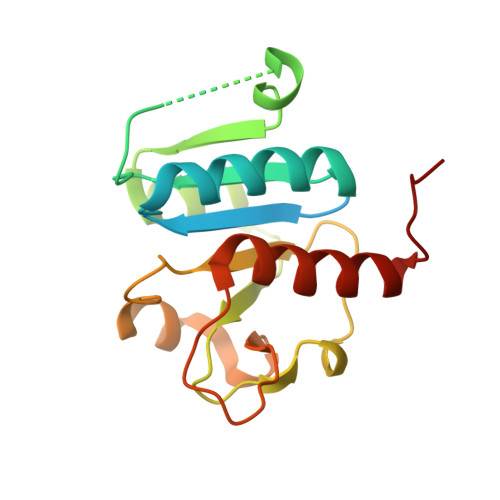 | |
UniProt | |||||
Find proteins for Q8E8X2 (Shewanella oneidensis (strain ATCC 700550 / JCM 31522 / CIP 106686 / LMG 19005 / NCIMB 14063 / MR-1)) Explore Q8E8X2 Go to UniProtKB: Q8E8X2 | |||||
Entity Groups | |||||
| Sequence Clusters | 30% Identity50% Identity70% Identity90% Identity95% Identity100% Identity | ||||
| UniProt Group | Q8E8X2 | ||||
Sequence AnnotationsExpand | |||||
| |||||
| Length ( Å ) | Angle ( ˚ ) |
|---|---|
| a = 74.816 | α = 90 |
| b = 77.988 | β = 90 |
| c = 101.044 | γ = 90 |
| Software Name | Purpose |
|---|---|
| REFMAC | refinement |
| HKL-2000 | data scaling |
| PDB_EXTRACT | data extraction |
| HKL-2000 | data reduction |
| MOLREP | phasing |
| Funding Organization | Location | Grant Number |
|---|---|---|
| National Research Foundation (NRF, Korea) | Korea, Republic Of | -- |


