7DD0

Crystal structure of the N-terminal domain of TagH from Bacillus subtilis


Experimental Data Snapshot

  • Method: X-RAY DIFFRACTION
  • Resolution: 2.70 Å
  • R-Value Free: 0.235 
  • R-Value Work: 0.186 
  • R-Value Observed: 0.189 

wwPDB Validation   3D Report Full Report


This is version 1.2 of the entry. See complete history


Literature

Crystal structure of the N-terminal domain of TagH reveals a potential drug targeting site.

Yang, C.S.Huang, W.C.Ko, T.P.Wang, Y.C.Wang, A.H.Chen, Y.

(2020) Biochem Biophys Res Commun 536: 1-6

  • DOI: https://doi.org/10.1016/j.bbrc.2020.12.028
  • Primary Citation of Related Structures:  
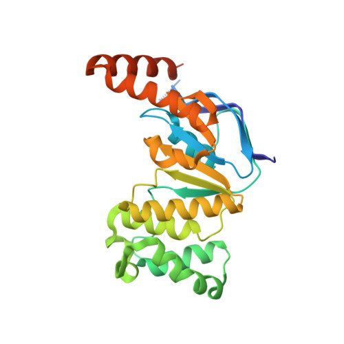
    7DD0

  • PubMed Abstract: 

    Bacterial wall teichoic acids (WTAs) are synthesized intracellularly and exported by a two-component transporter, TagGH, comprising the transmembrane and ATPase subunits TagG and TagH. Here the dimeric structure of the N-terminal domain of TagH (TagH-N) was solved by single-wavelength anomalous diffraction using a selenomethionine-containing crystal, which shows an ATP-binding cassette (ABC) architecture with RecA-like and helical subdomains. Besides significant structural differences from other ABC transporters, a prominent patch of positively charged surface is seen in the center of the TagH-N dimer, suggesting a potential binding site for the glycerol phosphate chain of WTA. The ATPase activity of TagH-N was inhibited by clodronate, a bisphosphonate, in a non-competitive manner, consistent with the proposed WTA-binding site for drug targeting.


  • Organizational Affiliation

    Institute of New Drug Development, China Medical University, Taichung, 406, Taiwan.


Macromolecules
Find similar proteins by:  (by identity cutoff)  |  3D Structure
Entity ID: 1
MoleculeChains Sequence LengthOrganismDetailsImage
Teichoic acids export ATP-binding protein TagH
A, B, C, D
280Bacillus subtilis subsp. subtilis str. 168Mutation(s): 0 
Gene Names: tagHBSU35700
EC: 7.5.2.4
UniProt
Find proteins for P42954 (Bacillus subtilis (strain 168))
Explore P42954 
Go to UniProtKB:  P42954
Entity Groups  
Sequence Clusters30% Identity50% Identity70% Identity90% Identity95% Identity100% Identity
UniProt GroupP42954
Sequence Annotations
Expand
  • Reference Sequence
Experimental Data & Validation

Experimental Data

  • Method: X-RAY DIFFRACTION
  • Resolution: 2.70 Å
  • R-Value Free: 0.235 
  • R-Value Work: 0.186 
  • R-Value Observed: 0.189 
  • Space Group: P 1 21 1
Unit Cell:
Length ( Å )Angle ( ˚ )
a = 73.735α = 90
b = 90.667β = 91.766
c = 91.213γ = 90
Software Package:
Software NamePurpose
PHENIXrefinement
HKL-2000data reduction
HKL-2000data scaling
AutoSolphasing

Structure Validation

View Full Validation Report


Entry History & Funding Information

Deposition Data


Funding OrganizationLocationGrant Number
Ministry of Science and Technology (MoST, Taiwan)Taiwan108-2311-B241-001

Revision History  (Full details and data files)

  • Version 1.0: 2020-12-23
    Type: Initial release
  • Version 1.1: 2021-01-13
    Changes: Database references
  • Version 1.2: 2024-03-27
    Changes: Data collection, Database references, Derived calculations, Refinement description