Experimental phasing with vanadium and application to nucleotide-binding membrane proteins.
El Omari, K., Mohamad, N., Bountra, K., Duman, R., Romano, M., Schlegel, K., Kwong, H.S., Mykhaylyk, V., Olesen, C., Moller, J.V., Bublitz, M., Beis, K., Wagner, A.(2020) IUCrJ 7: 1092-1101
- PubMed: 33209320
- DOI: https://doi.org/10.1107/S2052252520012312
- Primary Citation of Related Structures:
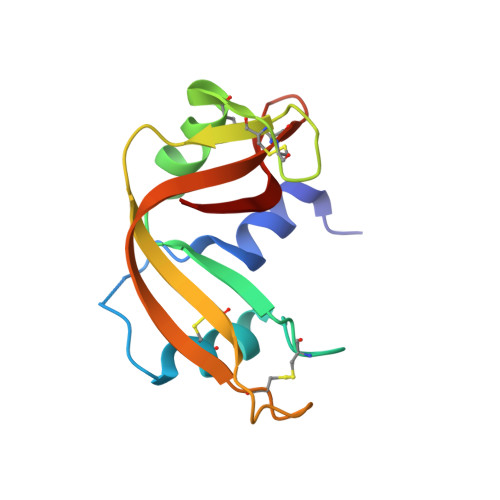
6YO1, 6YSO - PubMed Abstract:
The structure determination of soluble and membrane proteins can be hindered by the crystallographic phase problem, especially in the absence of a suitable homologous structure. Experimental phasing is the method of choice for novel structures; however, it often requires heavy-atom derivatization, which can be difficult and time-consuming. Here, a novel and rapid method to obtain experimental phases for protein structure determination by vanadium phasing is reported. Vanadate is a transition-state mimic of phosphoryl-transfer reactions and it has the advantage of binding specifically to the active site of numerous enzymes catalyzing this reaction. The applicability of vanadium phasing has been validated by determining the structures of three different protein-vanadium complexes, two of which are integral membrane proteins: the rabbit sarcoplasmic reticulum Ca 2+ -ATPase, the antibacterial peptide ATP-binding cassette transporter McjD from Escherichia coli and the soluble enzyme RNAse A from Bos taurus . Vanadium phasing was successful even at low resolution and despite severe anisotropy in the data. This method is principally applicable to a large number of proteins, representing six of the seven Enzyme Commission classes. It relies exclusively on the specific chemistry of the protein and it does not require any modifications, making it a very powerful addition to the phasing toolkit. In addition to the phasing power of this technique, the protein-vanadium complexes also provide detailed insights into the reaction mechanisms of the studied proteins.
- Diamond Light Source, Harwell Science and Innovation Campus, Didcot OX11 0DE, United Kingdom.
Organizational Affiliation: