Misannotations of the genes encoding sugar N-formyltransferases.
Girardi, N.M., Thoden, J.B., Holden, H.M.(2020) Protein Sci 29: 930-940
- PubMed: 31867814
- DOI: https://doi.org/10.1002/pro.3807
- Primary Citation of Related Structures:
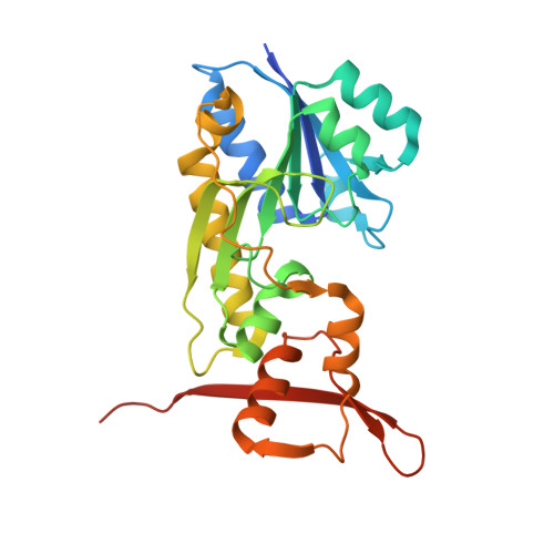
6V2T, 6V33 - PubMed Abstract:
Tens of thousands of bacterial genome sequences are now known due to the development of rapid and inexpensive sequencing technologies. An important key in utilizing these vast amounts of data in a biologically meaningful way is to infer the function of the proteins encoded in the genomes via bioinformatics techniques. Whereas these approaches are absolutely critical to the annotation of gene function, there are still issues of misidentifications, which must be experimentally corrected. For example, many of the bacterial DNA sequences encoding sugar N-formyltransferases have been annotated as l-methionyl-tRNA transferases in the databases. These mistakes may be due in part to the fact that until recently the structures and functions of these enzymes were not well known. Herein we describe the misannotation of two genes, WP_088211966.1 and WP_096244125.1, from Shewanella spp. and Pseudomonas congelans, respectively. Although the proteins encoded by these genes were originally suggested to function as l-methionyl-tRNA transferases, we demonstrate that they actually catalyze the conversion of dTDP-4-amino-4,6-dideoxy-d-glucose to dTDP-4-formamido-4,6-dideoxy-d-glucose utilizing N 10 -formyltetrahydrofolate as the carbon source. For this analysis, the genes encoding these enzymes were cloned and the corresponding proteins purified. X-ray structures of the two proteins were determined to high resolution and kinetic analyses were conducted. Both enzymes display classical Michaelis-Menten kinetics and adopt the characteristic three-dimensional structural fold previously observed for other sugar N-formyltransferases. The results presented herein will aid in the future annotation of these fascinating enzymes.
- Department of Biochemistry, University of Wisconsin, Madison, Wisconsin.
Organizational Affiliation: