6UZ0

Cardiac sodium channel with flecainide


Experimental Data Snapshot

  • Method: ELECTRON MICROSCOPY
  • Resolution: 3.24 Å
  • Aggregation State: PARTICLE 
  • Reconstruction Method: SINGLE PARTICLE 

wwPDB Validation   3D Report Full Report


This is version 2.0 of the entry. See complete history


Literature

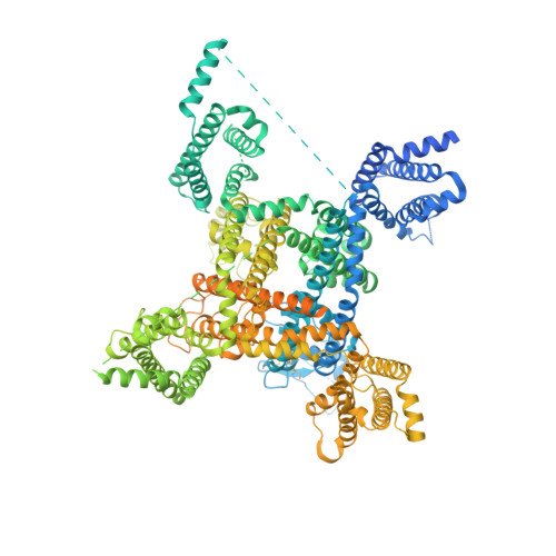
Structure of the Cardiac Sodium Channel.

Jiang, D.Shi, H.Tonggu, L.Gamal El-Din, T.M.Lenaeus, M.J.Zhao, Y.Yoshioka, C.Zheng, N.Catterall, W.A.

(2020) Cell 180: 122-134.e10

  • DOI: https://doi.org/10.1016/j.cell.2019.11.041
  • Primary Citation of Related Structures:  
    6UZ0, 6UZ3

  • PubMed Abstract: 

    Voltage-gated sodium channel Na v 1.5 generates cardiac action potentials and initiates the heartbeat. Here, we report structures of Na V 1.5 at 3.2-3.5 Å resolution. Na V 1.5 is distinguished from other sodium channels by a unique glycosyl moiety and loss of disulfide-bonding capability at the Na V β subunit-interaction sites. The antiarrhythmic drug flecainide specifically targets the central cavity of the pore. The voltage sensors are partially activated, and the fast-inactivation gate is partially closed. Activation of the voltage sensor of Domain III allows binding of the isoleucine-phenylalanine-methionine (IFM) motif to the inactivation-gate receptor. Asp and Ala, in the selectivity motif DEKA, line the walls of the ion-selectivity filter, whereas Glu and Lys are in positions to accept and release Na + ions via a charge-delocalization network. Arrhythmia mutation sites undergo large translocations during gating, providing a potential mechanism for pathogenic effects. Our results provide detailed insights into Na v 1.5 structure, pharmacology, activation, inactivation, ion selectivity, and arrhythmias.


  • Organizational Affiliation

    Department of Pharmacology, University of Washington, Seattle, WA 98195, USA.


Macromolecules
Find similar proteins by:  (by identity cutoff)  |  3D Structure
Entity ID: 1
MoleculeChains Sequence LengthOrganismDetailsImage
Sodium channel protein type 5 subunit alpha,Green fluorescent protein1,838Rattus norvegicusAequorea victoria
This entity is chimeric
Mutation(s): 0 
Gene Names: Scn5aGFP
Membrane Entity: Yes 
UniProt
Find proteins for P42212 (Aequorea victoria)
Explore P42212 
Go to UniProtKB:  P42212
Find proteins for P15389 (Rattus norvegicus)
Explore P15389 
Go to UniProtKB:  P15389
Entity Groups  
Sequence Clusters30% Identity50% Identity70% Identity90% Identity95% Identity100% Identity
UniProt GroupsP42212P15389
Sequence Annotations
Expand
  • Reference Sequence
Oligosaccharides

Help

Entity ID: 2
MoleculeChains Length2D Diagram Glycosylation3D Interactions
2-acetamido-2-deoxy-beta-D-glucopyranose-(1-4)-2-acetamido-2-deoxy-beta-D-glucopyranose
B, D
2N-Glycosylation
Glycosylation Resources
GlyTouCan:  G42666HT
GlyCosmos:  G42666HT
GlyGen:  G42666HT
Entity ID: 3
MoleculeChains Length2D Diagram Glycosylation3D Interactions
beta-D-mannopyranose-(1-4)-2-acetamido-2-deoxy-beta-D-glucopyranose-(1-4)-2-acetamido-2-deoxy-beta-D-glucopyranose
C
3N-Glycosylation
Glycosylation Resources
GlyTouCan:  G15407YE
GlyCosmos:  G15407YE
GlyGen:  G15407YE
Small Molecules
Ligands 4 Unique
IDChains Name / Formula / InChI Key2D Diagram3D Interactions
6OU
Query on 6OU

Download Ideal Coordinates CCD File 
L [auth A]
M [auth A]
N [auth A]
O [auth A]
P [auth A]
L [auth A],
M [auth A],
N [auth A],
O [auth A],
P [auth A],
Q [auth A],
R [auth A],
S [auth A],
T [auth A],
U [auth A],
V [auth A]
[(2~{R})-1-[2-azanylethoxy(oxidanyl)phosphoryl]oxy-3-hexadecanoyloxy-propan-2-yl] (~{Z})-octadec-9-enoate
C39 H76 N O8 P
FHQVHHIBKUMWTI-OTMQOFQLSA-N
9Z9
Query on 9Z9

Download Ideal Coordinates CCD File 
G [auth A],
H [auth A],
I [auth A],
J [auth A],
K [auth A]
(3beta,14beta,17beta,25R)-3-[4-methoxy-3-(methoxymethyl)butoxy]spirost-5-en
C34 H56 O5
CEEBZAXXSRFQIC-GZSGZGDASA-N
K4D (Subject of Investigation/LOI)
Query on K4D

Download Ideal Coordinates CCD File 
W [auth A]Flecainide
C17 H20 F6 N2 O3
DJBNUMBKLMJRSA-LLVKDONJSA-N
NAG
Query on NAG

Download Ideal Coordinates CCD File 
E [auth A],
F [auth A]
2-acetamido-2-deoxy-beta-D-glucopyranose
C8 H15 N O6
OVRNDRQMDRJTHS-FMDGEEDCSA-N
Experimental Data & Validation

Experimental Data

  • Method: ELECTRON MICROSCOPY
  • Resolution: 3.24 Å
  • Aggregation State: PARTICLE 
  • Reconstruction Method: SINGLE PARTICLE 
EM Software:
TaskSoftware PackageVersion
MODEL REFINEMENTPHENIX
RECONSTRUCTIONcisTEM

Structure Validation

View Full Validation Report


Entry History & Funding Information

Deposition Data


Funding OrganizationLocationGrant Number
National Institutes of Health/National Heart, Lung, and Blood Institute (NIH/NHLBI)United StatesR01 HL112808
Howard Hughes Medical Institute (HHMI)United States--

Revision History  (Full details and data files)

  • Version 1.0: 2020-01-01
    Type: Initial release
  • Version 1.1: 2020-01-08
    Changes: Database references
  • Version 1.2: 2020-01-22
    Changes: Database references
  • Version 2.0: 2020-07-29
    Type: Remediation
    Reason: Carbohydrate remediation
    Changes: Advisory, Atomic model, Data collection, Derived calculations, Structure summary