6EDS

Structure of Cysteine-free Human Insulin-Degrading Enzyme in complex with Glucagon and Substrate-selective Macrocyclic Inhibitor 63


Experimental Data Snapshot

  • Method: X-RAY DIFFRACTION
  • Resolution: 3.18 Å
  • R-Value Free: 0.222 
  • R-Value Work: 0.177 
  • R-Value Observed: 0.179 

wwPDB Validation   3D Report Full Report


Ligand Structure Quality Assessment 


This is version 1.3 of the entry. See complete history


Literature

Substrate-selective inhibitors that reprogram the activity of insulin-degrading enzyme.

Maianti, J.P.Tan, G.A.Vetere, A.Welsh, A.J.Wagner, B.K.Seeliger, M.A.Liu, D.R.

(2019) Nat Chem Biol 15: 565-574

  • DOI: https://doi.org/10.1038/s41589-019-0271-0
  • Primary Citation of Related Structures:  
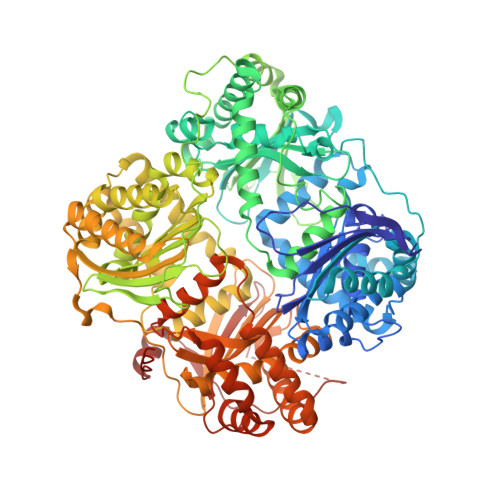
    6BYZ, 6EDS, 6MQ3

  • PubMed Abstract: 

    Enzymes that act on multiple substrates are common in biology but pose unique challenges as therapeutic targets. The metalloprotease insulin-degrading enzyme (IDE) modulates blood glucose levels by cleaving insulin, a hormone that promotes glucose clearance. However, IDE also degrades glucagon, a hormone that elevates glucose levels and opposes the effect of insulin. IDE inhibitors to treat diabetes, therefore, should prevent IDE-mediated insulin degradation, but not glucagon degradation, in contrast with traditional modes of enzyme inhibition. Using a high-throughput screen for non-active-site ligands, we discovered potent and highly specific small-molecule inhibitors that alter IDE's substrate selectivity. X-ray co-crystal structures, including an IDE-ligand-glucagon ternary complex, revealed substrate-dependent interactions that enable these inhibitors to potently block insulin binding while allowing glucagon cleavage, even at saturating inhibitor concentrations. These findings suggest a path for developing IDE-targeting therapeutics, and offer a blueprint for modulating other enzymes in a substrate-selective manner to unlock their therapeutic potential.


  • Organizational Affiliation

    Merkin Institute of Transformative Technologies in Healthcare, Broad Institute of MIT and Harvard, Cambridge, MA, USA.


Macromolecules
Find similar proteins by:  (by identity cutoff)  |  3D Structure
Entity ID: 1
MoleculeChains Sequence LengthOrganismDetailsImage
Insulin-degrading enzyme
A, B
978Homo sapiensMutation(s): 14 
Gene Names: IDE
EC: 3.4.24.56
UniProt & NIH Common Fund Data Resources
Find proteins for P14735 (Homo sapiens)
Explore P14735 
Go to UniProtKB:  P14735
PHAROS:  P14735
GTEx:  ENSG00000119912 
Entity Groups  
Sequence Clusters30% Identity50% Identity70% Identity90% Identity95% Identity100% Identity
UniProt GroupP14735
Sequence Annotations
Expand
  • Reference Sequence
Find similar proteins by:  (by identity cutoff)  |  3D Structure
Entity ID: 2
MoleculeChains Sequence LengthOrganismDetailsImage
Glucagon
C, D
29Homo sapiensMutation(s): 0 
Gene Names: GCG
UniProt & NIH Common Fund Data Resources
Find proteins for P01275 (Homo sapiens)
Explore P01275 
Go to UniProtKB:  P01275
PHAROS:  P01275
GTEx:  ENSG00000115263 
Entity Groups  
Sequence Clusters30% Identity50% Identity70% Identity90% Identity95% Identity100% Identity
UniProt GroupP01275
Sequence Annotations
Expand
  • Reference Sequence
Small Molecules
Ligands 4 Unique
IDChains Name / Formula / InChI Key2D Diagram3D Interactions
J22 (Subject of Investigation/LOI)
Query on J22

Download Ideal Coordinates CCD File 
E [auth A],
L [auth B]
{(8R,9S,10S)-9-(2',3'-dimethyl[1,1'-biphenyl]-4-yl)-6-[(1-methyl-1H-imidazol-2-yl)sulfonyl]-1,6-diazabicyclo[6.2.0]decan-10-yl}methanol
C27 H34 N4 O3 S
OJXBQVFJQWLDCX-NXCFDTQHSA-N
EPE
Query on EPE

Download Ideal Coordinates CCD File 
F [auth A],
I [auth B]
4-(2-HYDROXYETHYL)-1-PIPERAZINE ETHANESULFONIC ACID
C8 H18 N2 O4 S
JKMHFZQWWAIEOD-UHFFFAOYSA-N
PEG
Query on PEG

Download Ideal Coordinates CCD File 
G [auth A],
J [auth B]
DI(HYDROXYETHYL)ETHER
C4 H10 O3
MTHSVFCYNBDYFN-UHFFFAOYSA-N
DIO
Query on DIO

Download Ideal Coordinates CCD File 
H [auth A],
K [auth B]
1,4-DIETHYLENE DIOXIDE
C4 H8 O2
RYHBNJHYFVUHQT-UHFFFAOYSA-N
Experimental Data & Validation

Experimental Data

  • Method: X-RAY DIFFRACTION
  • Resolution: 3.18 Å
  • R-Value Free: 0.222 
  • R-Value Work: 0.177 
  • R-Value Observed: 0.179 
  • Space Group: P 65
Unit Cell:
Length ( Å )Angle ( ˚ )
a = 263.119α = 90
b = 263.119β = 90
c = 90.322γ = 120
Software Package:
Software NamePurpose
PHENIXrefinement
Cootmodel building
XDSdata reduction
autoPROCdata scaling
PHASERphasing

Structure Validation

View Full Validation Report


Ligand Structure Quality Assessment 


Entry History 

Deposition Data

Revision History  (Full details and data files)

  • Version 1.0: 2019-04-03
    Type: Initial release
  • Version 1.1: 2019-04-17
    Changes: Author supporting evidence, Data collection
  • Version 1.2: 2019-05-29
    Changes: Data collection, Database references
  • Version 1.3: 2023-10-11
    Changes: Data collection, Database references, Refinement description