5Y4D

Crystal Structure of AnkB Ankyrin Repeats in Complex with AnkR/AnkB Chimeric Autoinhibition Segment


Experimental Data Snapshot

  • Method: X-RAY DIFFRACTION
  • Resolution: 3.30 Å
  • R-Value Free: 0.229 
  • R-Value Work: 0.183 
  • R-Value Observed: 0.188 

wwPDB Validation   3D Report Full Report


This is version 1.1 of the entry. See complete history


Literature

Autoinhibition of ankyrin-B/G membrane target bindings by intrinsically disordered segments from the tail regions.

Chen, K.Li, J.Wang, C.Wei, Z.Zhang, M.

(2017) Elife 6

  • DOI: https://doi.org/10.7554/eLife.29150
  • Primary Citation of Related Structures:  
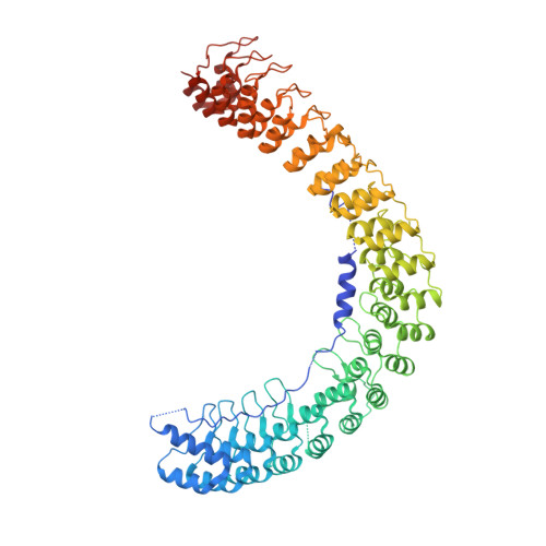
    5Y4D, 5Y4E, 5Y4F

  • PubMed Abstract: 

    Ankyrins together with their spectrin partners are the master organizers of micron-scale membrane domains in diverse tissues. The 24 ankyrin (ANK) repeats of ankyrins bind to numerous membrane proteins, linking them to spectrin-based cytoskeletons at specific membrane microdomains. The accessibility of the target binding groove of ANK repeats must be regulated to achieve spatially defined functions of ankyrins/target complexes in different tissues, though little is known in this regard. Here we systemically investigated the autoinhibition mechanism of ankyrin-B/G by combined biochemical, biophysical and structural biology approaches. We discovered that the entire ANK repeats are inhibited by combinatorial and quasi-independent bindings of multiple disordered segments located in the ankyrin-B/G linkers and tails, suggesting a mechanistic basis for differential regulations of membrane target bindings by ankyrins. In addition to elucidating the autoinhibition mechanisms of ankyrins, our study may also shed light on regulations on target bindings by other long repeat-containing proteins.


  • Organizational Affiliation

    Division of Life Science, State Key Laboratory of Molecular Neuroscience, Hong Kong University of Science and Technology, Hong Kong, China.


Macromolecules
Find similar proteins by:  (by identity cutoff)  |  3D Structure
Entity ID: 1
MoleculeChains Sequence LengthOrganismDetailsImage
Ankyrin-1,Ankyrin-2,Ankyrin-2731Mus musculusHomo sapiensMutation(s): 0 
Gene Names: Ank1ANK2
UniProt & NIH Common Fund Data Resources
Find proteins for Q02357 (Mus musculus)
Explore Q02357 
Go to UniProtKB:  Q02357
Find proteins for Q01484 (Homo sapiens)
Explore Q01484 
Go to UniProtKB:  Q01484
PHAROS:  Q01484
GTEx:  ENSG00000145362 
Entity Groups  
Sequence Clusters30% Identity50% Identity70% Identity90% Identity95% Identity100% Identity
UniProt GroupsQ02357Q01484
Sequence Annotations
Expand
  • Reference Sequence
Experimental Data & Validation

Experimental Data

  • Method: X-RAY DIFFRACTION
  • Resolution: 3.30 Å
  • R-Value Free: 0.229 
  • R-Value Work: 0.183 
  • R-Value Observed: 0.188 
  • Space Group: H 3 2
Unit Cell:
Length ( Å )Angle ( ˚ )
a = 179.787α = 90
b = 179.787β = 90
c = 227.098γ = 120
Software Package:
Software NamePurpose
SCALEPACKdata scaling
PHENIXrefinement
PDB_EXTRACTdata extraction
PHASERphasing

Structure Validation

View Full Validation Report


Entry History 

Deposition Data

Revision History  (Full details and data files)

  • Version 1.0: 2017-09-13
    Type: Initial release
  • Version 1.1: 2023-11-22
    Changes: Data collection, Database references, Refinement description