5TDV

Intermediate O2 diiron complex in the Q228A variant of Toluene 4-moonoxygenase (T4moHD)


Experimental Data Snapshot

  • Method: X-RAY DIFFRACTION
  • Resolution: 2.00 Å
  • R-Value Free: 0.195 
  • R-Value Work: 0.153 
  • R-Value Observed: 0.155 

wwPDB Validation   3D Report Full Report


This is version 1.5 of the entry. See complete history


Literature

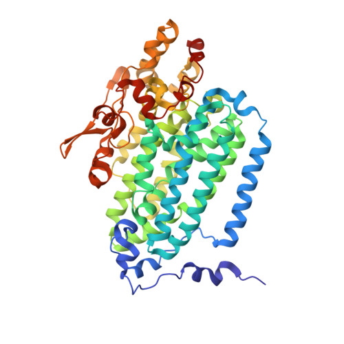
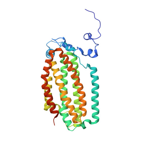
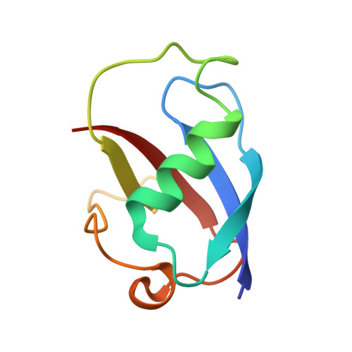
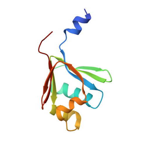
In-crystal reaction cycle of a toluene-bound diiron hydroxylase.

Acheson, J.F.Bailey, L.J.Brunold, T.C.Fox, B.G.

(2017) Nature 544: 191-195

  • DOI: https://doi.org/10.1038/nature21681
  • Primary Citation of Related Structures:  
    5TDS, 5TDT, 5TDU, 5TDV

  • PubMed Abstract: 

    Electrophilic aromatic substitution is one of the most important and recognizable classes of organic chemical transformation. Enzymes create the strong electrophiles that are needed for these highly energetic reactions by using O 2 , electrons, and metals or other cofactors. Although the nature of the oxidants that carry out electrophilic aromatic substitution has been deduced from many approaches, it has been difficult to determine their structures. Here we show the structure of a diiron hydroxylase intermediate formed during a reaction with toluene. Density functional theory geometry optimizations of an active site model reveal that the intermediate is an arylperoxo Fe 2+ /Fe 3+ species with delocalized aryl radical character. The structure suggests that a carboxylate ligand of the diiron centre may trigger homolytic cleavage of the O-O bond by transferring a proton from a metal-bound water. Our work provides the spatial and electronic constraints needed to propose a comprehensive mechanism for diiron enzyme arene hydroxylation that accounts for many prior experimental results.


  • Organizational Affiliation

    Department of Biochemistry, University of Wisconsin-Madison, Madison, Wisconsin 53706, USA.


Macromolecules
Find similar proteins by:  (by identity cutoff)  |  3D Structure
Entity ID: 1
MoleculeChains Sequence LengthOrganismDetailsImage
Toluene-4-monooxygenase system protein AA,
E [auth D]
500Pseudomonas mendocinaMutation(s): 1 
Gene Names: tmoA
EC: 1.14.13
UniProt
Find proteins for Q00456 (Pseudomonas mendocina)
Explore Q00456 
Go to UniProtKB:  Q00456
Entity Groups  
Sequence Clusters30% Identity50% Identity70% Identity90% Identity95% Identity100% Identity
UniProt GroupQ00456
Sequence Annotations
Expand
  • Reference Sequence
Find similar proteins by:  (by identity cutoff)  |  3D Structure
Entity ID: 2
MoleculeChains Sequence LengthOrganismDetailsImage
Toluene-4-monooxygenase system protein E
B, F
327Pseudomonas mendocinaMutation(s): 0 
Gene Names: tmoE
EC: 1.14.13
UniProt
Find proteins for Q00460 (Pseudomonas mendocina)
Explore Q00460 
Go to UniProtKB:  Q00460
Entity Groups  
Sequence Clusters30% Identity50% Identity70% Identity90% Identity95% Identity100% Identity
UniProt GroupQ00460
Sequence Annotations
Expand
  • Reference Sequence
Find similar proteins by:  (by identity cutoff)  |  3D Structure
Entity ID: 3
MoleculeChains Sequence LengthOrganismDetailsImage
Toluene-4-monooxygenase system protein B
C, G
84Pseudomonas mendocinaMutation(s): 0 
Gene Names: tmoB
EC: 1.14.13
UniProt
Find proteins for Q00457 (Pseudomonas mendocina)
Explore Q00457 
Go to UniProtKB:  Q00457
Entity Groups  
Sequence Clusters30% Identity50% Identity70% Identity90% Identity95% Identity100% Identity
UniProt GroupQ00457
Sequence Annotations
Expand
  • Reference Sequence
Find similar proteins by:  (by identity cutoff)  |  3D Structure
Entity ID: 4
MoleculeChains Sequence LengthOrganismDetailsImage
Toluene-4-monooxygenase system protein DD [auth E],
H
103Pseudomonas mendocinaMutation(s): 0 
Gene Names: tmoD
EC: 1.14.13
UniProt
Find proteins for Q00459 (Pseudomonas mendocina)
Explore Q00459 
Go to UniProtKB:  Q00459
Entity Groups  
Sequence Clusters30% Identity50% Identity70% Identity90% Identity95% Identity100% Identity
UniProt GroupQ00459
Sequence Annotations
Expand
  • Reference Sequence
Experimental Data & Validation

Experimental Data

  • Method: X-RAY DIFFRACTION
  • Resolution: 2.00 Å
  • R-Value Free: 0.195 
  • R-Value Work: 0.153 
  • R-Value Observed: 0.155 
  • Space Group: P 21 21 21
Unit Cell:
Length ( Å )Angle ( ˚ )
a = 100.293α = 90
b = 115.757β = 90
c = 181.654γ = 90
Software Package:
Software NamePurpose
PHENIXrefinement
HKL-2000data reduction
HKL-2000data scaling
MOLREPphasing

Structure Validation

View Full Validation Report


Entry History & Funding Information

Deposition Data


Funding OrganizationLocationGrant Number
National Science Foundation (NSF, United States)United StatesMCB-0843239
Department of Energy (DOE, United States)United StatesW-31-109-ENG-38

Revision History  (Full details and data files)

  • Version 1.0: 2017-03-22
    Type: Initial release
  • Version 1.1: 2017-04-12
    Changes: Database references
  • Version 1.2: 2017-04-26
    Changes: Database references
  • Version 1.3: 2017-09-20
    Changes: Author supporting evidence
  • Version 1.4: 2019-11-27
    Changes: Author supporting evidence
  • Version 1.5: 2023-10-04
    Changes: Data collection, Database references, Derived calculations, Refinement description