5KB3

1.4 A resolution structure of Helicobacter Pylori MTAN in complexed with p-ClPh-DADMe-ImmA

  • Classification: HYDROLASE
  • Organism(s): Helicobacter pylori J99
  • Expression System: Escherichia coli
  • Mutation(s): No 

  • Deposited: 2016-06-02 Released: 2016-11-16 
  • Deposition Author(s): Banco, M.T., Ronning, D.R.
  • Funding Organization(s): National Aeronautic Space Administration (NASA, United States), National Institutes of Health/National Institute Of Allergy and Infectious Diseases (NIH/NIAID)

Experimental Data Snapshot

  • Method: X-RAY DIFFRACTION
  • Resolution: 1.40 Å
  • R-Value Free: 0.170 
  • R-Value Work: 0.139 
  • R-Value Observed: 0.140 

wwPDB Validation   3D Report Full Report


Ligand Structure Quality Assessment 


This is version 2.2 of the entry. See complete history


Literature

Neutron structures of the Helicobacter pylori 5'-methylthioadenosine nucleosidase highlight proton sharing and protonation states.

Banco, M.T.Mishra, V.Ostermann, A.Schrader, T.E.Evans, G.B.Kovalevsky, A.Ronning, D.R.

(2016) Proc Natl Acad Sci U S A 113: 13756-13761

  • DOI: https://doi.org/10.1073/pnas.1609718113
  • Primary Citation of Related Structures:  
    5CCD, 5CCE, 5JPC, 5K1Z, 5KB3

  • PubMed Abstract: 

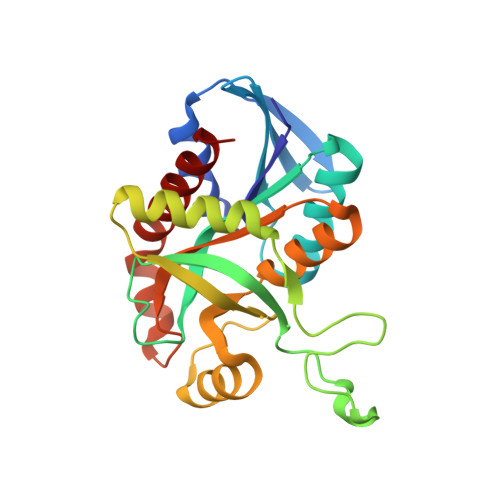
    MTAN (5'-methylthioadenosine nucleosidase) catalyzes the hydrolysis of the N-ribosidic bond of a variety of adenosine-containing metabolites. The Helicobacter pylori MTAN (HpMTAN) hydrolyzes 6-amino-6-deoxyfutalosine in the second step of the alternative menaquinone biosynthetic pathway. Substrate binding of the adenine moiety is mediated almost exclusively by hydrogen bonds, and the proposed catalytic mechanism requires multiple proton-transfer events. Of particular interest is the protonation state of residue D198, which possesses a pK a above 8 and functions as a general acid to initiate the enzymatic reaction. In this study we present three corefined neutron/X-ray crystal structures of wild-type HpMTAN cocrystallized with S-adenosylhomocysteine (SAH), Formycin A (FMA), and (3R,4S)-4-(4-Chlorophenylthiomethyl)-1-[(9-deaza-adenin-9-yl)methyl]-3-hydroxypyrrolidine (p-ClPh-Thio-DADMe-ImmA) as well as one neutron/X-ray crystal structure of an inactive variant (HpMTAN-D198N) cocrystallized with SAH. These results support a mechanism of D198 pKa elevation through the unexpected sharing of a proton with atom N7 of the adenine moiety possessing unconventional hydrogen-bond geometry. Additionally, the neutron structures also highlight active site features that promote the stabilization of the transition state and slight variations in these interactions that result in 100-fold difference in binding affinities between the DADMe-ImmA and ImmA analogs.


  • Organizational Affiliation

    Department of Chemistry and Biochemistry, University of Toledo, Toledo, OH 43606.


Macromolecules
Find similar proteins by:  (by identity cutoff)  |  3D Structure
Entity ID: 1
MoleculeChains Sequence LengthOrganismDetailsImage
Aminodeoxyfutalosine nucleosidase230Helicobacter pylori J99Mutation(s): 0 
Gene Names: mtnNmtnjhp_0082
EC: 3.2.2.30 (PDB Primary Data), 3.2.2.9 (PDB Primary Data)
UniProt
Find proteins for Q9ZMY2 (Helicobacter pylori (strain J99 / ATCC 700824))
Explore Q9ZMY2 
Go to UniProtKB:  Q9ZMY2
Entity Groups  
Sequence Clusters30% Identity50% Identity70% Identity90% Identity95% Identity100% Identity
UniProt GroupQ9ZMY2
Sequence Annotations
Expand
  • Reference Sequence
Small Molecules
Ligands 2 Unique
IDChains Name / Formula / InChI Key2D Diagram3D Interactions
4CT
Query on 4CT

Download Ideal Coordinates CCD File 
B [auth A](3R,4S)-1-[(4-amino-5H-pyrrolo[3,2-d]pyrimidin-7-yl)methyl]-4-{[(4-chlorophenyl)sulfanyl]methyl}pyrrolidin-3-ol
C18 H20 Cl N5 O S
MZZBHZOHYGEGEE-DOMZBBRYSA-N
MG
Query on MG

Download Ideal Coordinates CCD File 
C [auth A]MAGNESIUM ION
Mg
JLVVSXFLKOJNIY-UHFFFAOYSA-N
Binding Affinity Annotations 
IDSourceBinding Affinity
4CT BindingDB:  5KB3 Kd: 0.57 (nM) from 1 assay(s)
Binding MOAD:  5KB3 Kd: 0.57 (nM) from 1 assay(s)
Experimental Data & Validation

Experimental Data

  • Method: X-RAY DIFFRACTION
  • Resolution: 1.40 Å
  • R-Value Free: 0.170 
  • R-Value Work: 0.139 
  • R-Value Observed: 0.140 
  • Space Group: P 32 2 1
Unit Cell:
Length ( Å )Angle ( ˚ )
a = 81.207α = 90
b = 81.207β = 90
c = 67.254γ = 120
Software Package:
Software NamePurpose
PHENIXrefinement
HKL-2000data reduction
SCALEPACKdata scaling
PDB_EXTRACTdata extraction

Structure Validation

View Full Validation Report


Ligand Structure Quality Assessment 


Entry History & Funding Information

Deposition Data


Funding OrganizationLocationGrant Number
National Aeronautic Space Administration (NASA, United States)United StatesN-123528-01
National Institutes of Health/National Institute Of Allergy and Infectious Diseases (NIH/NIAID)United StatesAI105084

Revision History  (Full details and data files)

  • Version 1.0: 2016-11-16
    Type: Initial release
  • Version 1.1: 2016-12-21
    Changes: Database references
  • Version 1.2: 2017-09-13
    Changes: Author supporting evidence
  • Version 2.0: 2019-12-11
    Changes: Atomic model, Author supporting evidence
  • Version 2.1: 2020-01-01
    Changes: Author supporting evidence
  • Version 2.2: 2023-09-27
    Changes: Data collection, Database references, Derived calculations, Refinement description