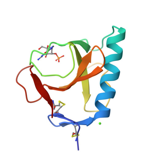
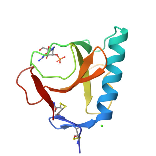
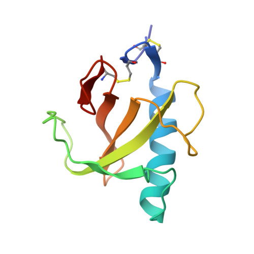
Conserved water molecules in a large family of microbial ribonucleases.
Loris, R., Langhorst, U., De Vos, S., Decanniere, K., Bouckaert, J., Maes, D., Transue, T.R., Steyaert, J.(1999) Proteins 36: 117-134
- PubMed: 10373011
- DOI: https://doi.org/10.1002/(sici)1097-0134(19990701)36:1<117::aid-prot10>3.0.co;2-h
- Primary Citation of Related Structures:
1BU4, 2BU4, 3BU4, 4BU4, 5BU4 - PubMed Abstract:
We systematically analyzed the crystallographically determined water molecules of all known structures of RNase T1 and compared them to the ordered solvent in a large number of related microbial nucleases. To assess the crystallographers' impact on the interpretation of the solvent structure, we independently refined five validation structures from diffraction data derived from five isomorphous crystals of RNase T1. We also compared the positions of water molecules found in 11 published isomorphous RNase T1 inhibitor complexes. These data suggest that the positions of most of the waters located on the surface of a protein and that are well-determined in the experimental electron density maps are determined primarily by crystal packing forces. Water molecules with less well-defined electron density are in general unique to one or a small number of crystal structures. Only a small number of the well-defined waters are found to be independent of the crystal environment. These waters have a low accessible surface area and B-factor, and tend to be conserved in the crystal structures of a number of evolutionary related ribonucleases as well. A single water molecule is found conserved in all known microbial ribonucleases.
Organizational Affiliation:
Laboratorium voor Ultrastructuur, Vlaams Interuniversitair Instituut voor Biotechnologie, Vrije Universiteit Brussel, Sint-Genesius-Rode, Belgium. reloris@vub.ac.be


