5FXI

GluN1b-GluN2B NMDA receptor structure in non-active-2 conformation


Experimental Data Snapshot

  • Method: ELECTRON MICROSCOPY
  • Resolution: 6.40 Å
  • Aggregation State: PARTICLE 
  • Reconstruction Method: SINGLE PARTICLE 

wwPDB Validation   3D Report Full Report


This is version 1.4 of the entry. See complete history


Literature

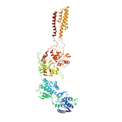
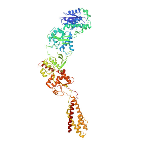
Activation of Nmda Receptors and the Mechanism of Inhibition by Ifenprodil.

Tajima, N.Karakas, E.Grant, T.Simorowski, N.Diaz-Avalos, R.Grigorieff, N.Furukawa, H.

(2016) Nature 534: 63

  • DOI: https://doi.org/10.1038/nature17679
  • Primary Citation of Related Structures:  
    5B3J, 5FXG, 5FXH, 5FXI, 5FXJ, 5FXK

  • PubMed Abstract: 

    The physiology of N-methyl-d-aspartate (NMDA) receptors is fundamental to brain development and function. NMDA receptors are ionotropic glutamate receptors that function as heterotetramers composed mainly of GluN1 and GluN2 subunits. Activation of NMDA receptors requires binding of neurotransmitter agonists to a ligand-binding domain (LBD) and structural rearrangement of an amino-terminal domain (ATD). Recent crystal structures of GluN1-GluN2B NMDA receptors bound to agonists and an allosteric inhibitor, ifenprodil, represent the allosterically inhibited state. However, how the ATD and LBD move to activate the NMDA receptor ion channel remains unclear. Here we applied X-ray crystallography, single-particle electron cryomicroscopy and electrophysiology to rat NMDA receptors to show that, in the absence of ifenprodil, the bi-lobed structure of GluN2 ATD adopts an open conformation accompanied by rearrangement of the GluN1-GluN2 ATD heterodimeric interface, altering subunit orientation in the ATD and LBD and forming an active receptor conformation that gates the ion channel.


  • Organizational Affiliation

    Cold Spring Harbor Laboratory, W. M. Keck Structural Biology Laboratory, Cold Spring Harbor, New York 11724, USA.


Macromolecules
Find similar proteins by:  (by identity cutoff)  |  3D Structure
Entity ID: 1
MoleculeChains Sequence LengthOrganismDetailsImage
N-METHYL-D-ASPARTATE RECEPTOR GLUN1
A, C
846Rattus norvegicusMutation(s): 14 
Membrane Entity: Yes 
UniProt
Find proteins for P35439 (Rattus norvegicus)
Explore P35439 
Go to UniProtKB:  P35439
Entity Groups  
Sequence Clusters30% Identity50% Identity70% Identity90% Identity95% Identity100% Identity
UniProt GroupP35439
Sequence Annotations
Expand
  • Reference Sequence
Find similar proteins by:  (by identity cutoff)  |  3D Structure
Entity ID: 2
MoleculeChains Sequence LengthOrganismDetailsImage
N-METHYL-D-ASPARTATE RECEPTOR GLUN2B
B, D
827Rattus norvegicusMutation(s): 5 
Membrane Entity: Yes 
UniProt
Find proteins for Q00960 (Rattus norvegicus)
Explore Q00960 
Go to UniProtKB:  Q00960
Entity Groups  
Sequence Clusters30% Identity50% Identity70% Identity90% Identity95% Identity100% Identity
UniProt GroupQ00960
Sequence Annotations
Expand
  • Reference Sequence
Experimental Data & Validation

Experimental Data

  • Method: ELECTRON MICROSCOPY
  • Resolution: 6.40 Å
  • Aggregation State: PARTICLE 
  • Reconstruction Method: SINGLE PARTICLE 
EM Software:
TaskSoftware PackageVersion
RECONSTRUCTIONFREALIGN

Structure Validation

View Full Validation Report


Entry History 

Deposition Data

Revision History  (Full details and data files)

  • Version 1.0: 2016-05-11
    Type: Initial release
  • Version 1.1: 2016-06-15
    Changes: Database references
  • Version 1.2: 2017-04-19
    Changes: Other
  • Version 1.3: 2017-08-23
    Changes: Data collection
  • Version 1.4: 2019-12-18
    Changes: Other