4Y7S

Crystal Structure of the CFEM protein Csa2


Experimental Data Snapshot

  • Method: X-RAY DIFFRACTION
  • Resolution: 2.00 Å
  • R-Value Free: 0.192 
  • R-Value Work: 0.153 
  • R-Value Observed: 0.154 

wwPDB Validation   3D Report Full Report


Ligand Structure Quality Assessment 


This is version 1.1 of the entry. See complete history


Literature

Structural basis of haem-iron acquisition by fungal pathogens.

Nasser, L.Weissman, Z.Pinsky, M.Amartely, H.Dvir, H.Kornitzer, D.

(2016) Nat Microbiol 1: 16156-16156

  • DOI: https://doi.org/10.1038/nmicrobiol.2016.156
  • Primary Citation of Related Structures:  
    4Y7S

  • PubMed Abstract: 

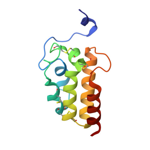
    Pathogenic microorganisms must cope with extremely low free-iron concentrations in the host's tissues. Some fungal pathogens rely on secreted haemophores that belong to the Common in Fungal Extracellular Membrane (CFEM) protein family, to extract haem from haemoglobin and to transfer it to the cell's interior, where it can serve as a source of iron. Here we report the first three-dimensional structure of a CFEM protein, the haemophore Csa2 secreted by Candida albicans. The CFEM domain adopts a novel helical-basket fold that consists of six α-helices, and is uniquely stabilized by four disulfide bonds formed by its eight signature cysteines. The planar haem molecule is bound between a flat hydrophobic platform located on top of the helical basket and a peripheral N-terminal 'handle' extension. Exceptionally, an aspartic residue serves as the CFEM axial ligand, and so confers coordination of Fe 3+ haem, but not of Fe 2+ haem. Histidine substitution mutants of this conserved Asp acquired Fe 2+ haem binding and retained the capacity to extract haem from haemoglobin. However, His-substituted CFEM proteins were not functional in vivo and showed disturbed haem exchange in vitro, which suggests a role for the oxidation-state-specific Asp coordination in haem acquisition by CFEM proteins.


  • Organizational Affiliation

    B. Rappaport Faculty of Medicine, Technion - Israel Institute of Technology, and the Rappaport Institute for Research in the Medical Sciences, Haifa 31096, Israel.


Macromolecules
Find similar proteins by:  (by identity cutoff)  |  3D Structure
Entity ID: 1
MoleculeChains Sequence LengthOrganismDetailsImage
Surface antigen protein 2
A, B, C
111Candida albicansMutation(s): 0 
UniProt
Find proteins for Q5A0X8 (Candida albicans (strain SC5314 / ATCC MYA-2876))
Explore Q5A0X8 
Go to UniProtKB:  Q5A0X8
Entity Groups  
Sequence Clusters30% Identity50% Identity70% Identity90% Identity95% Identity100% Identity
UniProt GroupQ5A0X8
Sequence Annotations
Expand
  • Reference Sequence
Experimental Data & Validation

Experimental Data

  • Method: X-RAY DIFFRACTION
  • Resolution: 2.00 Å
  • R-Value Free: 0.192 
  • R-Value Work: 0.153 
  • R-Value Observed: 0.154 
  • Space Group: P 21 21 21
Unit Cell:
Length ( Å )Angle ( ˚ )
a = 61.341α = 90
b = 61.208β = 90
c = 97.3γ = 90
Software Package:
Software NamePurpose
HKL-3000data reduction
SCALEPACKdata scaling
SHELXphasing
MLPHAREphasing
DMphasing
PHENIXrefinement
PDB_EXTRACTdata extraction

Structure Validation

View Full Validation Report


Ligand Structure Quality Assessment 


Entry History & Funding Information

Deposition Data


Funding OrganizationLocationGrant Number
Marie CurieIsrael330879
Israeli Ministry of Health's Chief Scientist OfficeIsrael--
Russell Berrie Nanotechnology Institute - TechnionIsrael--
Lorry I. Lokey Interdiciplinary Center for Life Sciences and EngineeringIsrael--

Revision History  (Full details and data files)

  • Version 1.0: 2016-08-03
    Type: Initial release
  • Version 1.1: 2016-10-12
    Changes: Database references