4WHJ

Myxovirus Resistance Protein 2 (MxB)


Experimental Data Snapshot

  • Method: X-RAY DIFFRACTION
  • Resolution: 3.20 Å
  • R-Value Free: 0.299 
  • R-Value Work: 0.265 
  • R-Value Observed: 0.267 

wwPDB Validation   3D Report Full Report


This is version 1.3 of the entry. See complete history


Literature

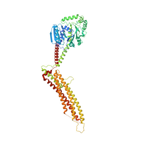
Structural Insight into HIV-1 Restriction by MxB.

Fribourgh, J.L.Nguyen, H.C.Matreyek, K.A.Alvarez, F.J.Summers, B.J.Dewdney, T.G.Aiken, C.Zhang, P.Engelman, A.Xiong, Y.

(2014) Cell Host Microbe 16: 627-638

  • DOI: https://doi.org/10.1016/j.chom.2014.09.021
  • Primary Citation of Related Structures:  
    4WHJ

  • PubMed Abstract: 

    The myxovirus resistance (Mx) proteins are interferon-induced dynamin GTPases that can inhibit a variety of viruses. Recently, MxB, but not MxA, was shown to restrict HIV-1 by an unknown mechanism that likely occurs in close proximity to the host cell nucleus and involves the viral capsid. Here, we present the crystal structure of MxB and reveal determinants involved in HIV-1 restriction. MxB adopts an extended antiparallel dimer and dimerization, but not higher-ordered oligomerization, is critical for restriction. Although MxB is structurally similar to MxA, the orientation of individual domains differs between MxA and MxB, and their antiviral functions rely on separate determinants, indicating distinct mechanisms for virus inhibition. Additionally, MxB directly binds the HIV-1 capsid, and this interaction depends on dimerization and the N terminus of MxB as well as the assembled capsid lattice. These insights establish a framework for understanding the mechanism by which MxB restricts HIV-1.


  • Organizational Affiliation

    Yale University, Molecular Biophysics and Biochemistry, New Haven, CT 06520, USA.


Macromolecules
Find similar proteins by:  (by identity cutoff)  |  3D Structure
Entity ID: 1
MoleculeChains Sequence LengthOrganismDetailsImage
Interferon-induced GTP-binding protein Mx2
A, B
646Homo sapiensMutation(s): 4 
Gene Names: MX2
UniProt & NIH Common Fund Data Resources
Find proteins for P20592 (Homo sapiens)
Explore P20592 
Go to UniProtKB:  P20592
PHAROS:  P20592
GTEx:  ENSG00000183486 
Entity Groups  
Sequence Clusters30% Identity50% Identity70% Identity90% Identity95% Identity100% Identity
UniProt GroupP20592
Sequence Annotations
Expand
  • Reference Sequence
Experimental Data & Validation

Experimental Data

  • Method: X-RAY DIFFRACTION
  • Resolution: 3.20 Å
  • R-Value Free: 0.299 
  • R-Value Work: 0.265 
  • R-Value Observed: 0.267 
  • Space Group: P 1 21 1
Unit Cell:
Length ( Å )Angle ( ˚ )
a = 53.185α = 90
b = 80.782β = 95.73
c = 183.666γ = 90
Software Package:
Software NamePurpose
PHENIXrefinement
HKL-2000data reduction
PDB_EXTRACTdata extraction
SCALEPACKdata scaling
DENZOdata reduction

Structure Validation

View Full Validation Report


Entry History & Funding Information

Deposition Data


Funding OrganizationLocationGrant Number
Collaboration Development Pilot Program award from the Pittsburgh Center for HIV Protein InteractionsUnited States--

Revision History  (Full details and data files)

  • Version 1.0: 2014-10-29
    Type: Initial release
  • Version 1.1: 2014-12-10
    Changes: Database references
  • Version 1.2: 2017-11-22
    Changes: Derived calculations, Other, Refinement description, Source and taxonomy
  • Version 1.3: 2023-09-27
    Changes: Data collection, Database references, Refinement description