


Mechanism and function of lipid-directed oligomerization of vinculin
Chinthalapudi, K., Rangarajan, E.S., Patil, D., Izard, T.To be published.
Experimental Data Snapshot
Starting Model: experimental
View more details
Entity ID: 1 | |||||
|---|---|---|---|---|---|
| Molecule | Chains | Sequence Length | Organism | Details | Image |
| Vinculin | 176 | Homo sapiens | Mutation(s): 1 Gene Names: VCL | 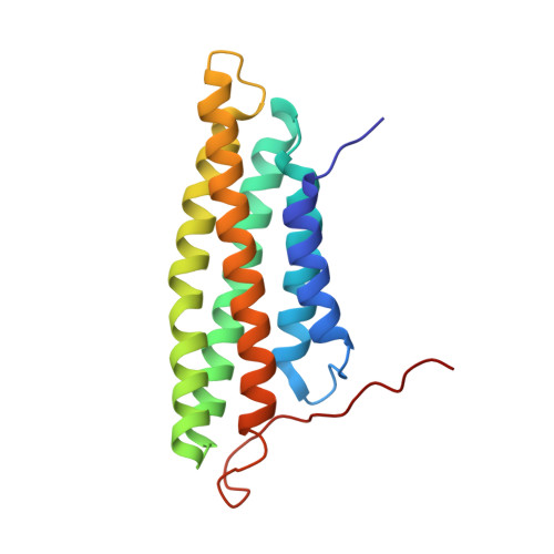 | |
UniProt & NIH Common Fund Data Resources | |||||
Find proteins for P18206 (Homo sapiens) Explore P18206 Go to UniProtKB: P18206 | |||||
PHAROS: P18206 GTEx: ENSG00000035403 | |||||
Entity Groups | |||||
| Sequence Clusters | 30% Identity50% Identity70% Identity90% Identity95% Identity100% Identity | ||||
| UniProt Group | P18206 | ||||
Sequence AnnotationsExpand | |||||
| |||||
| Ligands 2 Unique | |||||
|---|---|---|---|---|---|
| ID | Chains | Name / Formula / InChI Key | 2D Diagram | 3D Interactions | |
| PIO Query on PIO | G [auth A], H [auth B], K [auth E] | [(2R)-2-octanoyloxy-3-[oxidanyl-[(1R,2R,3S,4R,5R,6S)-2,3,6-tris(oxidanyl)-4,5-diphosphonooxy-cyclohexyl]oxy-phosphoryl]oxy-propyl] octanoate C25 H49 O19 P3 XLNCEHRXXWQMPK-MJUMVPIBSA-N |  | ||
| GOL Query on GOL | I [auth B], J [auth C], L [auth F] | GLYCEROL C3 H8 O3 PEDCQBHIVMGVHV-UHFFFAOYSA-N |  | ||
| Length ( Å ) | Angle ( ˚ ) |
|---|---|
| a = 102.579 | α = 90 |
| b = 102.579 | β = 90 |
| c = 190.772 | γ = 120 |
| Software Name | Purpose |
|---|---|
| HKL-2000 | data collection |
| MOLREP | phasing |
| BUSTER | refinement |
| XDS | data reduction |
| autoPROC | data scaling |
| SCALA | data scaling |


