4MLI

Crystal structure of the SpyTag/SpyCatcher complex


Experimental Data Snapshot

  • Method: X-RAY DIFFRACTION
  • Resolution: 2.10 Å
  • R-Value Free: 0.225 
  • R-Value Work: 0.198 
  • R-Value Observed: 0.200 

wwPDB Validation   3D Report Full Report


This is version 1.2 of the entry. See complete history


Literature

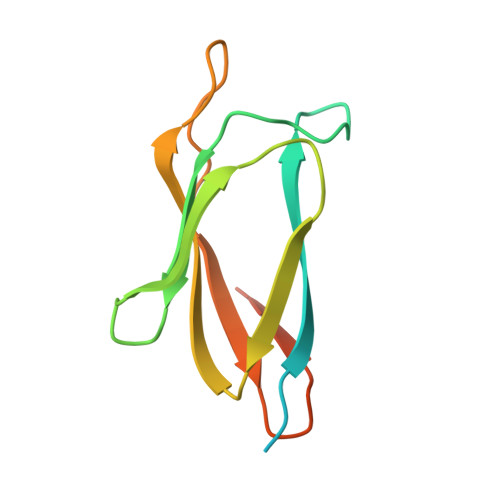
Structural Analysis and Optimization of the Covalent Association between SpyCatcher and a Peptide Tag.

Li, L.Fierer, J.O.Rapoport, T.A.Howarth, M.

(2014) J Mol Biol 426: 309-317

  • DOI: https://doi.org/10.1016/j.jmb.2013.10.021
  • Primary Citation of Related Structures:  
    4MLI, 4MLS

  • PubMed Abstract: 

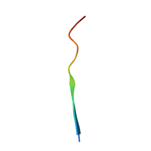
    Peptide tagging is a key strategy for observing and isolating proteins. However, the interactions of proteins with peptides are nearly all rapidly reversible. Proteins tagged with the peptide SpyTag form an irreversible covalent bond to the SpyCatcher protein via a spontaneous isopeptide linkage, thereby offering a genetically encoded way to create peptide interactions that resist force and harsh conditions. Here, we determined the crystal structure of the reconstituted covalent complex of SpyTag and SpyCatcher at 2.1Å resolution. The structure showed the expected reformation of the β-sandwich domain seen in the parental streptococcal adhesin, but flanking sequences at both N- and C-termini of SpyCatcher were disordered. In addition, only 10 out of 13 amino acids of the SpyTag peptide were observed to interact with SpyCatcher, pointing to specific contacts important for rapid split protein reconstitution. Based on these structural insights, we expressed a range of SpyCatcher variants and identified a minimized SpyCatcher, 32 residues shorter, that maintained rapid reaction with SpyTag. Together, these results give insight into split protein β-strand complementation and enhance a distinct approach to ultrastable molecular interaction.


  • Organizational Affiliation

    Howard Hughes Medical Institute and Department of Cell Biology, Harvard Medical School, 240 Longwood Avenue, Boston, MA 02115, USA.


Macromolecules
Find similar proteins by:  (by identity cutoff)  |  3D Structure
Entity ID: 1
MoleculeChains Sequence LengthOrganismDetailsImage
Fibronectin binding proteinA,
B [auth C]
116Streptococcus pyogenesMutation(s): 2 
Gene Names: CnaB2fba2
UniProt
Find proteins for Q8G9G1 (Streptococcus pyogenes)
Explore Q8G9G1 
Go to UniProtKB:  Q8G9G1
Entity Groups  
Sequence Clusters30% Identity50% Identity70% Identity90% Identity95% Identity100% Identity
UniProt GroupQ8G9G1
Sequence Annotations
Expand
  • Reference Sequence

Find similar proteins by:  Sequence   |   3D Structure  

Entity ID: 2
MoleculeChains Sequence LengthOrganismDetailsImage
SpyTagC [auth B],
D
13N/AMutation(s): 0 
Entity Groups  
Sequence Clusters30% Identity50% Identity70% Identity90% Identity95% Identity100% Identity
Sequence Annotations
Expand
  • Reference Sequence
Experimental Data & Validation

Experimental Data

  • Method: X-RAY DIFFRACTION
  • Resolution: 2.10 Å
  • R-Value Free: 0.225 
  • R-Value Work: 0.198 
  • R-Value Observed: 0.200 
  • Space Group: P 1
  • Diffraction Data: https://doi.org/10.15785/SBGRID/90
Unit Cell:
Length ( Å )Angle ( ˚ )
a = 31.621α = 83.7
b = 38.527β = 78.25
c = 44.491γ = 89.72
Software Package:
Software NamePurpose
HKL-2000data collection
PHASERphasing
PHENIXrefinement
HKL-2000data reduction
HKL-2000data scaling

Structure Validation

View Full Validation Report


Entry History 

Deposition Data

Revision History  (Full details and data files)

  • Version 1.0: 2013-11-13
    Type: Initial release
  • Version 1.1: 2014-01-22
    Changes: Database references
  • Version 1.2: 2023-09-20
    Changes: Data collection, Database references, Derived calculations, Refinement description