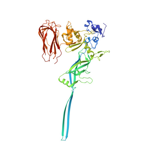
44BV | pdb_000044bv

Iota toxin Ib D452A pore


Experimental Data Snapshot

  • Method: ELECTRON MICROSCOPY
  • Resolution: 2.56 Å
  • Aggregation State: PARTICLE 
  • Reconstruction Method: SINGLE PARTICLE 

Starting Model: experimental
View more details

wwPDB Validation 3D Report Full Report

Validation slider image for 44BV

This is version 1.1 of the entry. See complete history. 

Literature

Step-by-step maturation mechanism of the binary toxin pore revealed by cryo-EM analysis.

Yamada, T., Nakanishi, R., Sugita, Y., Ninomiya, Y., Yoshida, T., Noda, T., Tsuge, H.

(2026) Proc Natl Acad Sci U S A 123: e2627537123-e2627537123

  • DOI: https://doi.org/10.1073/pnas.2627537123
  • Primary Citation Related Structures: 
    44BV, 44BW

  • PubMed Abstract: 

    Membrane pore-forming proteins (PFPs) form ring-shaped membrane-translocating oligomers on membranes, contributing to infection, immunity, and cell death functions. Binary toxins produced by some bacteria consist of an enzymatic component that acts as a toxin and a membrane-binding component that forms a pore that delivers the enzymatic component into target cells. Cryoelectron microscopy (cryo-EM) has advanced our understanding of these translocation mechanisms by revealing several binary toxin complexes' structures. However, the mechanisms underlying the initial pore formation remain unclear. We determined the structures of several oligomeric forms of the membrane-binding component Ib of the iota toxin from Clostridium perfringens at various stages of pore formation. Structural comparisons revealed how the symmetrically arranged soluble oligomer (prepore) asymmetrically matures into a transmembrane oligomer (pore). These findings enhance our understanding of mechanisms of PFP and provide a structural basis for developing nanodevices using membrane pores.


  • Organizational Affiliation: 
    • Department of Frontier Life Sciences, Faculty of Life Sciences, Kyoto Sangyo University, Kyoto 603-8555, Japan.

Macromolecule Content 

  • Total Structure Weight: 410.23 kDa 
  • Atom Count: 26,957 
  • Modeled Residue Count: 3,451 
  • Deposited Residue Count: 3,710 
  • Unique protein chains: 1

Macromolecules

Find similar proteins by:|  3D Structure
Entity ID: 1
MoleculeChains  Sequence LengthOrganismDetailsImage
Iota toxin component Ib
A, B, C, D, E
A, B, C, D, E, F, G
530Clostridium perfringensMutation(s): 1 
UniProt
Find proteins for Q46221 (Clostridium perfringens)
Explore Q46221 
Go to UniProtKB:  Q46221
Entity Groups
Sequence Clusters30% Identity50% Identity70% Identity90% Identity95% Identity100% Identity
UniProt GroupQ46221
Sequence Annotations
Expand
Reference Sequence

Small Molecules

Ligands 1 Unique
IDChains Name / Formula / InChI Key2D Diagram3D Interactions
CA
(Subject of Investigation/LOI)

Query on CA


Download:Ideal Coordinates CCD File
AA [auth G]
BA [auth G]
H [auth A]
I [auth A]
J [auth A]
AA [auth G],
BA [auth G],
H [auth A],
I [auth A],
J [auth A],
K [auth B],
L [auth B],
M [auth B],
N [auth C],
O [auth C],
P [auth C],
Q [auth D],
R [auth D],
S [auth D],
T [auth E],
U [auth E],
V [auth E],
W [auth F],
X [auth F],
Y [auth F],
Z [auth G]
CALCIUM ION
Ca
BHPQYMZQTOCNFJ-UHFFFAOYSA-N

Experimental Data & Validation

Experimental Data

  • Method: ELECTRON MICROSCOPY
  • Resolution: 2.56 Å
  • Aggregation State: PARTICLE 
  • Reconstruction Method: SINGLE PARTICLE 
EM Software:
TaskSoftware PackageVersion
RECONSTRUCTIONcryoSPARCv.4.5.3
MODEL REFINEMENTPHENIX2.0-5936
MODEL REFINEMENTCoot0.9.5

Structure Validation

View Full Validation Report


Entry History 

& Funding Information

Deposition Data


Funding OrganizationLocationGrant Number
Japan Society for the Promotion of Science (JSPS)Japan21J13410
Japan Society for the Promotion of Science (JSPS)Japan21H02452
Japan Society for the Promotion of Science (JSPS)Japan24K01993
Japan Agency for Medical Research and Development (AMED)Japan2366
Japan Agency for Medical Research and Development (AMED)Japan6392

Revision History  (Full details and data files)

  • Version 1.0: 2026-08-12
    Type: Initial release
  • Version 1.1: 2026-10-14
    Changes: Data collection, Database references