3QMZ

Crystal structure of the cytoplasmic dynein heavy chain motor domain


Experimental Data Snapshot

  • Method: X-RAY DIFFRACTION
  • Resolution: 6.00 Å
  • R-Value Free: 0.430 
  • R-Value Work: 0.430 

wwPDB Validation   3D Report Full Report


This is version 1.3 of the entry. See complete history


Literature

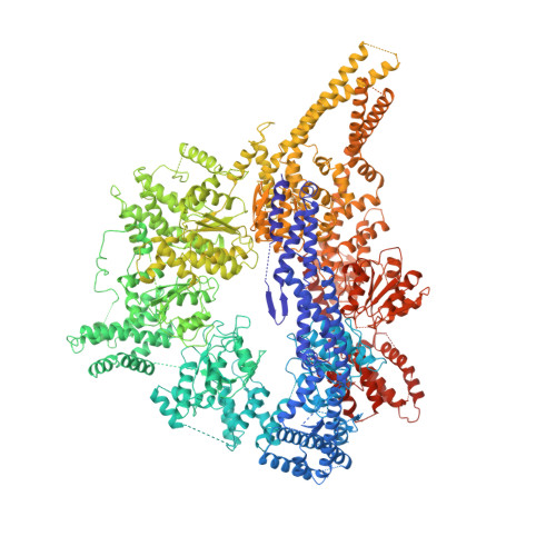
Crystal structure of the dynein motor domain.

Carter, A.P.Cho, C.Jin, L.Vale, R.D.

(2011) Science 331: 1159-1165

  • DOI: https://doi.org/10.1126/science.1202393
  • Primary Citation of Related Structures:  
    3QMZ

  • PubMed Abstract: 

    Dyneins are microtubule-based motor proteins that power ciliary beating, transport intracellular cargos, and help to construct the mitotic spindle. Evolved from ring-shaped hexameric AAA-family adenosine triphosphatases (ATPases), dynein's large size and complexity have posed challenges for understanding its structure and mechanism. Here, we present a 6 angstrom crystal structure of a functional dimer of two ~300-kilodalton motor domains of yeast cytoplasmic dynein. The structure reveals an unusual asymmetric arrangement of ATPase domains in the ring-shaped motor domain, the manner in which the mechanical element interacts with the ATPase ring, and an unexpected interaction between two coiled coils that create a base for the microtubule binding domain. The arrangement of these elements provides clues as to how adenosine triphosphate-driven conformational changes might be transmitted across the motor domain.


  • Organizational Affiliation

    Department of Cellular and Molecular Pharmacology, Howard Hughes Medical Institute, University of California-San Francisco, 600 16th Street, San Francisco, CA 94158, USA. cartera@mrc-lmb.cam.ac.uk


Macromolecules
Find similar proteins by:  (by identity cutoff)  |  3D Structure
Entity ID: 1
MoleculeChains Sequence LengthOrganismDetailsImage
Cytoplasmic dynein heavy chain
A, B
2,486Saccharomyces cerevisiaeMutation(s): 0 
UniProt
Find proteins for P36022 (Saccharomyces cerevisiae (strain ATCC 204508 / S288c))
Explore P36022 
Go to UniProtKB:  P36022
Entity Groups  
Sequence Clusters30% Identity50% Identity70% Identity90% Identity95% Identity100% Identity
UniProt GroupP36022
Sequence Annotations
Expand
  • Reference Sequence
Find similar proteins by:  (by identity cutoff)  |  3D Structure
Entity ID: 2
MoleculeChains Sequence LengthOrganismDetailsImage
Glutathione-S-transferaseC [auth T],
D [auth S]
219Schistosoma japonicumMutation(s): 0 
UniProt
Find proteins for P08515 (Schistosoma japonicum)
Explore P08515 
Go to UniProtKB:  P08515
Entity Groups  
Sequence Clusters30% Identity50% Identity70% Identity90% Identity95% Identity100% Identity
UniProt GroupP08515
Sequence Annotations
Expand
  • Reference Sequence
Experimental Data & Validation

Experimental Data

  • Method: X-RAY DIFFRACTION
  • Resolution: 6.00 Å
  • R-Value Free: 0.430 
  • R-Value Work: 0.430 
  • Space Group: P 1 21 1
Unit Cell:
Length ( Å )Angle ( ˚ )
a = 174.08α = 90
b = 118.921β = 90.27
c = 200.509γ = 90
Software Package:
Software NamePurpose
HKL-2000data collection
PHENIXmodel building
CNSrefinement
HKL-2000data reduction
HKL-2000data scaling
PHENIXphasing

Structure Validation

View Full Validation Report


Entry History 

Deposition Data

Revision History  (Full details and data files)

  • Version 1.0: 2011-03-16
    Type: Initial release
  • Version 1.1: 2011-07-13
    Changes: Version format compliance
  • Version 1.2: 2012-02-29
    Changes: Database references
  • Version 1.3: 2024-02-21
    Changes: Data collection, Database references