3OD2

E. coli NikR soaked with excess nickel ions


Experimental Data Snapshot

  • Method: X-RAY DIFFRACTION
  • Resolution: 2.60 Å
  • R-Value Free: 0.286 
  • R-Value Work: 0.236 

wwPDB Validation   3D Report Full Report


Ligand Structure Quality Assessment 


This is version 1.2 of the entry. See complete history


Literature

Structural basis of low-affinity nickel binding to the nickel-responsive transcription factor NikR from Escherichia coli.

Phillips, C.M.Schreiter, E.R.Stultz, C.M.Drennan, C.L.

(2010) Biochemistry 49: 7830-7838

  • DOI: https://doi.org/10.1021/bi100923j
  • Primary Citation of Related Structures:  
    3OD2

  • PubMed Abstract: 

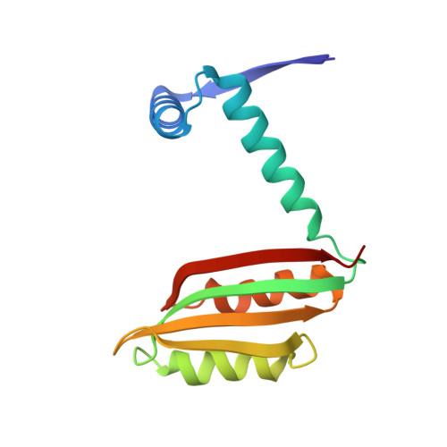
    Escherichia coli NikR regulates cellular nickel uptake by binding to the nik operon in the presence of nickel and blocking transcription of genes encoding the nickel uptake transporter. NikR has two binding affinities for the nik operon: a nanomolar dissociation constant with stoichiometric nickel and a picomolar dissociation constant with excess nickel [Bloom, S. L., and Zamble, D. B. (2004) Biochemistry 43, 10029-10038; Chivers, P. T., and Sauer, R. T. (2002) Chem. Biol. 9, 1141-1148]. While it is known that the stoichiometric nickel ions bind at the NikR tetrameric interface [Schreiter, E. R., et al. (2003) Nat. Struct. Biol. 10, 794-799; Schreiter, E. R., et al. (2006) Proc. Natl. Acad. Sci. U.S.A. 103, 13676-13681], the binding sites for excess nickel ions have not been fully described. Here we have determined the crystal structure of NikR in the presence of excess nickel to 2.6 A resolution and have obtained nickel anomalous data (1.4845 A) in the presence of excess nickel for both NikR alone and NikR cocrystallized with a 30-nucleotide piece of double-stranded DNA containing the nik operon. These anomalous data show that excess nickel ions do not bind to a single location on NikR but instead reveal a total of 22 possible low-affinity nickel sites on the NikR tetramer. These sites, for which there are six different types, are all on the surface of NikR, and most are found in both the NikR alone and NikR-DNA structures. Using a combination of crystallographic data and molecular dynamics simulations, the nickel sites can be described as preferring octahedral geometry, utilizing one to three protein ligands (typically histidine) and at least two water molecules.


  • Organizational Affiliation

    Department of Chemistry, Massachusetts Institute of Technology,Cambridge, Massachusetts 02139, USA.


Macromolecules
Find similar proteins by:  (by identity cutoff)  |  3D Structure
Entity ID: 1
MoleculeChains Sequence LengthOrganismDetailsImage
Nickel-responsive regulatory protein
A, B
133Escherichia coliMutation(s): 0 
Gene Names: ECs4348
UniProt
Find proteins for P0A6Z6 (Escherichia coli (strain K12))
Explore P0A6Z6 
Go to UniProtKB:  P0A6Z6
Entity Groups  
Sequence Clusters30% Identity50% Identity70% Identity90% Identity95% Identity100% Identity
UniProt GroupP0A6Z6
Sequence Annotations
Expand
  • Reference Sequence
Small Molecules
Ligands 2 Unique
IDChains Name / Formula / InChI Key2D Diagram3D Interactions
3CM
Query on 3CM

Download Ideal Coordinates CCD File 
I [auth B]3-CYCLOHEXYLPROPYL 4-O-ALPHA-D-GLUCOPYRANOSYL-BETA-D-GLUCOPYRANOSIDE
C21 H38 O11
FDBLAHXBTQUZSM-ZESVGKPKSA-N
NI
Query on NI

Download Ideal Coordinates CCD File 
C [auth A]
D [auth A]
E [auth A]
F [auth A]
G [auth A]
C [auth A],
D [auth A],
E [auth A],
F [auth A],
G [auth A],
H [auth A],
J [auth B],
K [auth B],
L [auth B],
M [auth B],
N [auth B]
NICKEL (II) ION
Ni
VEQPNABPJHWNSG-UHFFFAOYSA-N
Experimental Data & Validation

Experimental Data

  • Method: X-RAY DIFFRACTION
  • Resolution: 2.60 Å
  • R-Value Free: 0.286 
  • R-Value Work: 0.236 
  • Space Group: P 31 2 1
Unit Cell:
Length ( Å )Angle ( ˚ )
a = 50.735α = 90
b = 50.735β = 90
c = 183.114γ = 120
Software Package:
Software NamePurpose
MAR345dtbdata collection
CNSrefinement
DENZOdata reduction
SCALEPACKdata scaling
CNSphasing

Structure Validation

View Full Validation Report


Ligand Structure Quality Assessment 


Entry History 

Deposition Data

Revision History  (Full details and data files)

  • Version 1.0: 2010-09-08
    Type: Initial release
  • Version 1.1: 2011-07-13
    Changes: Version format compliance
  • Version 1.2: 2023-09-06
    Changes: Data collection, Database references, Derived calculations, Refinement description