3O8E

Structure of extracelllar portion of CD46 in complex with Adenovirus type 11 knob


Experimental Data Snapshot

  • Method: X-RAY DIFFRACTION
  • Resolution: 2.84 Å
  • R-Value Free: 0.229 
  • R-Value Work: 0.210 
  • R-Value Observed: 0.210 

wwPDB Validation   3D Report Full Report


Ligand Structure Quality Assessment 


This is version 2.1 of the entry. See complete history


Literature

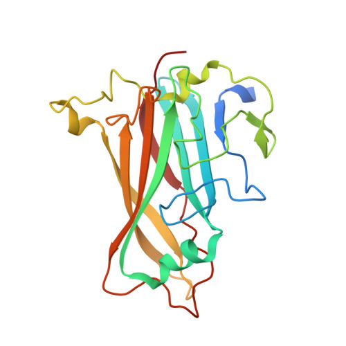
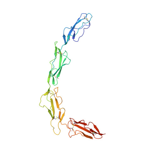
Structure of the extracellular portion of CD46 provides insights into its interactions with complement proteins and pathogens.

Persson, B.D.Schmitz, N.B.Santiago, C.Zocher, G.Larvie, M.Scheu, U.Casasnovas, J.M.Stehle, T.

(2010) PLoS Pathog 6: e1001122-e1001122

  • DOI: https://doi.org/10.1371/journal.ppat.1001122
  • Primary Citation of Related Structures:  
    3O8E

  • PubMed Abstract: 

    The human membrane cofactor protein (MCP, CD46) is a central component of the innate immune system. CD46 protects autologous cells from complement attack by binding to complement proteins C3b and C4b and serving as a cofactor for their cleavage. Recent data show that CD46 also plays a role in mediating acquired immune responses, and in triggering autophagy. In addition to these physiologic functions, a significant number of pathogens, including select adenoviruses, measles virus, human herpes virus 6 (HHV-6), Streptococci, and Neisseria, use CD46 as a cell attachment receptor. We have determined the crystal structure of the extracellular region of CD46 in complex with the human adenovirus type 11 fiber knob. Extracellular CD46 comprises four short consensus repeats (SCR1-SCR4) that form an elongated structure resembling a hockey stick, with a long shaft and a short blade. Domains SCR1, SCR2 and SCR3 are arranged in a nearly linear fashion. Unexpectedly, however, the structure reveals a profound bend between domains SCR3 and SCR4, which has implications for the interactions with ligands as well as the orientation of the protein at the cell surface. This bend can be attributed to an insertion of five hydrophobic residues in a SCR3 surface loop. Residues in this loop have been implicated in interactions with complement, indicating that the bend participates in binding to C3b and C4b. The structure provides an accurate framework for mapping all known ligand binding sites onto the surface of CD46, thereby advancing an understanding of how CD46 acts as a receptor for pathogens and physiologic ligands of the immune system.


  • Organizational Affiliation

    University of Tuebingen, Tuebingen, Germany.


Macromolecules
Find similar proteins by:  (by identity cutoff)  |  3D Structure
Entity ID: 1
MoleculeChains Sequence LengthOrganismDetailsImage
Fiber 36.1 kDa protein
A, C
213Human adenovirus 11Mutation(s): 0 
Gene Names: CD46MCPMIC10
UniProt
Find proteins for Q772X2 (Human adenovirus 11)
Explore Q772X2 
Go to UniProtKB:  Q772X2
Entity Groups  
Sequence Clusters30% Identity50% Identity70% Identity90% Identity95% Identity100% Identity
UniProt GroupQ772X2
Sequence Annotations
Expand
  • Reference Sequence
Find similar proteins by:  (by identity cutoff)  |  3D Structure
Entity ID: 2
MoleculeChains Sequence LengthOrganismDetailsImage
Membrane cofactor protein
B, D
252Homo sapiensMutation(s): 0 
UniProt & NIH Common Fund Data Resources
Find proteins for P15529 (Homo sapiens)
Explore P15529 
Go to UniProtKB:  P15529
PHAROS:  P15529
GTEx:  ENSG00000117335 
Entity Groups  
Sequence Clusters30% Identity50% Identity70% Identity90% Identity95% Identity100% Identity
UniProt GroupP15529
Sequence Annotations
Expand
  • Reference Sequence
Oligosaccharides

Help

Entity ID: 3
MoleculeChains Length2D Diagram Glycosylation3D Interactions
2-acetamido-2-deoxy-beta-D-glucopyranose-(1-4)-2-acetamido-2-deoxy-beta-D-glucopyranose
E, F, G
2N-Glycosylation
Glycosylation Resources
GlyTouCan:  G42666HT
GlyCosmos:  G42666HT
GlyGen:  G42666HT
Experimental Data & Validation

Experimental Data

  • Method: X-RAY DIFFRACTION
  • Resolution: 2.84 Å
  • R-Value Free: 0.229 
  • R-Value Work: 0.210 
  • R-Value Observed: 0.210 
  • Space Group: P 63
Unit Cell:
Length ( Å )Angle ( ˚ )
a = 108.16α = 90
b = 108.16β = 90
c = 222.99γ = 120
Software Package:
Software NamePurpose
PHASERphasing
BUSTERrefinement
XDSdata reduction
XDSdata scaling

Structure Validation

View Full Validation Report


Ligand Structure Quality Assessment 


Entry History 

Deposition Data

Revision History  (Full details and data files)

  • Version 1.0: 2010-10-13
    Type: Initial release
  • Version 1.1: 2011-07-13
    Changes: Version format compliance
  • Version 1.2: 2011-08-24
    Changes: Database references
  • Version 1.3: 2011-09-07
    Changes: Database references
  • Version 1.4: 2011-10-05
    Changes: Database references
  • Version 1.5: 2017-11-08
    Changes: Refinement description
  • Version 2.0: 2020-07-29
    Type: Remediation
    Reason: Carbohydrate remediation
    Changes: Atomic model, Data collection, Database references, Derived calculations, Structure summary
  • Version 2.1: 2023-09-06
    Changes: Data collection, Database references, Refinement description, Structure summary