3O6X

Crystal Structure of the type III Glutamine Synthetase from Bacteroides fragilis


Experimental Data Snapshot

  • Method: X-RAY DIFFRACTION
  • Resolution: 3.50 Å
  • R-Value Free: 0.269 
  • R-Value Work: 0.239 

wwPDB Validation   3D Report Full Report


Ligand Structure Quality Assessment 


This is version 1.2 of the entry. See complete history


Literature

Crystal Structure of Type III Glutamine Synthetase: Surprising Reversal of the Inter-Ring Interface.

van Rooyen, J.M.Abratt, V.R.Belrhali, H.Sewell, T.

(2011) Structure 19: 471-483

  • DOI: https://doi.org/10.1016/j.str.2011.02.001
  • Primary Citation of Related Structures:  
    3O6X

  • PubMed Abstract: 

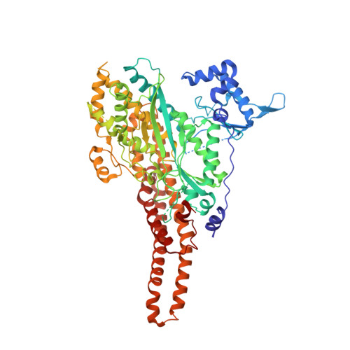
    Glutamine synthetases are ubiquitous, homo-oligomeric enzymes essential for nitrogen metabolism. Unlike types I and II, which are well described both structurally and functionally, the larger, type IIIs are poorly characterized despite their widespread occurrence. An understanding of the structural basis for this divergence and the implications for design of type-specific inhibitors has, therefore, been impossible. The first crystal structure of a GSIII enzyme, presented here, reveals a conservation of the GS catalytic fold but subtle differences in protein-ligand interactions suggest possible avenues for the design GSIII inhibitors. Despite these similarities, the divergence of the GSIII enzymes can be explained by differences in quaternary structure. Unexpectedly, the two hexameric rings of the GSIII dodecamer associate on the opposite surface relative to types I and II. The diversity of GS quaternary structures revealed here suggests a nonallosteric role for the evolution of the double-ringed architecture seen in all GS enzymes.


  • Organizational Affiliation

    Electron Microscope Unit, University of Cape Town, Rondebosch 7701, South Africa.


Macromolecules
Find similar proteins by:  (by identity cutoff)  |  3D Structure
Entity ID: 1
MoleculeChains Sequence LengthOrganismDetailsImage
Glutamine synthetase
A, B, C, D, E
A, B, C, D, E, F
729Bacteroides fragilis NCTC 9343Mutation(s): 0 
Gene Names: BF0955glnAGlnN
UniProt
Find proteins for Q5LGP1 (Bacteroides fragilis (strain ATCC 25285 / DSM 2151 / CCUG 4856 / JCM 11019 / NCTC 9343 / Onslow))
Explore Q5LGP1 
Go to UniProtKB:  Q5LGP1
Entity Groups  
Sequence Clusters30% Identity50% Identity70% Identity90% Identity95% Identity100% Identity
UniProt GroupQ5LGP1
Sequence Annotations
Expand
  • Reference Sequence
Small Molecules
Ligands 4 Unique
IDChains Name / Formula / InChI Key2D Diagram3D Interactions
ADP
Query on ADP

Download Ideal Coordinates CCD File 
CA [auth D]
IA [auth E]
J [auth A]
OA [auth F]
Q [auth B]
CA [auth D],
IA [auth E],
J [auth A],
OA [auth F],
Q [auth B],
W [auth C]
ADENOSINE-5'-DIPHOSPHATE
C10 H15 N5 O10 P2
XTWYTFMLZFPYCI-KQYNXXCUSA-N
P3S
Query on P3S

Download Ideal Coordinates CCD File 
BA [auth D]
HA [auth E]
I [auth A]
NA [auth F]
P [auth B]
BA [auth D],
HA [auth E],
I [auth A],
NA [auth F],
P [auth B],
V [auth C]
L-METHIONINE-S-SULFOXIMINE PHOSPHATE
C5 H13 N2 O6 P S
QQFOFBSCSWFFPB-NMAPHRJESA-N
CL
Query on CL

Download Ideal Coordinates CCD File 
DA [auth D]
EA [auth D]
JA [auth E]
K [auth A]
KA [auth E]
DA [auth D],
EA [auth D],
JA [auth E],
K [auth A],
KA [auth E],
L [auth A],
M [auth A],
PA [auth F],
R [auth B],
S [auth B],
X [auth C],
Y [auth C]
CHLORIDE ION
Cl
VEXZGXHMUGYJMC-UHFFFAOYSA-M
MG
Query on MG

Download Ideal Coordinates CCD File 
AA [auth D]
FA [auth E]
G [auth A]
GA [auth E]
H [auth A]
AA [auth D],
FA [auth E],
G [auth A],
GA [auth E],
H [auth A],
LA [auth F],
MA [auth F],
N [auth B],
O [auth B],
T [auth C],
U [auth C],
Z [auth D]
MAGNESIUM ION
Mg
JLVVSXFLKOJNIY-UHFFFAOYSA-N
Experimental Data & Validation

Experimental Data

  • Method: X-RAY DIFFRACTION
  • Resolution: 3.50 Å
  • R-Value Free: 0.269 
  • R-Value Work: 0.239 
  • Space Group: C 2 2 21
Unit Cell:
Length ( Å )Angle ( ˚ )
a = 198.25α = 90
b = 203.96β = 90
c = 234.59γ = 90
Software Package:
Software NamePurpose
MOSFLMdata reduction
SCALAdata scaling
CNSrefinement
PDB_EXTRACTdata extraction
PHENIXphasing

Structure Validation

View Full Validation Report


Ligand Structure Quality Assessment 


Entry History 

Deposition Data

Revision History  (Full details and data files)

  • Version 1.0: 2011-03-09
    Type: Initial release
  • Version 1.1: 2011-07-13
    Changes: Version format compliance
  • Version 1.2: 2024-02-21
    Changes: Data collection, Database references, Derived calculations