3MCY

Crystal structure of FimH lectin domain bound to biphenyl mannoside meta-methyl ester.


Experimental Data Snapshot

  • Method: X-RAY DIFFRACTION
  • Resolution: 2.90 Å
  • R-Value Free: 0.245 
  • R-Value Work: 0.219 
  • R-Value Observed: 0.220 

wwPDB Validation   3D Report Full Report


Ligand Structure Quality Assessment 


This is version 1.2 of the entry. See complete history


Literature

Structure-based drug design and optimization of mannoside bacterial FimH antagonists.

Han, Z.Pinkner, J.S.Ford, B.Obermann, R.Nolan, W.Wildman, S.A.Hobbs, D.Ellenberger, T.Cusumano, C.K.Hultgren, S.J.Janetka, J.W.

(2010) J Med Chem 53: 4779-4792

  • DOI: https://doi.org/10.1021/jm100438s
  • Primary Citation of Related Structures:  
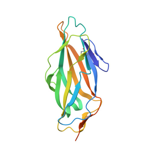
    3MCY

  • PubMed Abstract: 

    FimH-mediated cellular adhesion to mannosylated proteins is critical in the ability of uropathogenic E. coli (UPEC) to colonize and invade the bladder epithelium during urinary tract infection. We describe the discovery and optimization of potent small-molecule FimH bacterial adhesion antagonists based on alpha-d-mannose 1-position anomeric glycosides using X-ray structure-guided drug design. Optimized biarylmannosides display low nanomolar binding affinity for FimH in a fluorescence polarization assay and submicromolar cellular activity in a hemagglutination (HA) functional cell assay of bacterial adhesion. X-ray crystallography demonstrates that the biphenyl moiety makes several key interactions with the outer surface of FimH including pi-pi interactions with Tyr-48 and an H-bonding electrostatic interaction with the Arg-98/Glu-50 salt bridge. Dimeric analogues linked through the biaryl ring show an impressive 8-fold increase in potency relative to monomeric matched pairs and represent the most potent FimH antagonists identified to date. The FimH antagonists described herein hold great potential for development as novel therapeutics for the effective treatment of urinary tract infections.


  • Organizational Affiliation

    Department of Biochemistry and Molecular Biophysics, Washington University School of Medicine, 660 S. Euclid Avenue, St. Louis, Missouri 63110, USA.


Macromolecules
Find similar proteins by:  (by identity cutoff)  |  3D Structure
Entity ID: 1
MoleculeChains Sequence LengthOrganismDetailsImage
FimHA,
B [auth D],
C [auth B],
D [auth C]
181Escherichia coliMutation(s): 0 
Gene Names: fimH
UniProt
Find proteins for Q9S497 (Escherichia coli)
Explore Q9S497 
Go to UniProtKB:  Q9S497
Entity Groups  
Sequence Clusters30% Identity50% Identity70% Identity90% Identity95% Identity100% Identity
UniProt GroupQ9S497
Sequence Annotations
Expand
  • Reference Sequence
Small Molecules
Ligands 4 Unique
IDChains Name / Formula / InChI Key2D Diagram3D Interactions
ZH1
Query on ZH1

Download Ideal Coordinates CCD File 
E [auth A],
I [auth D],
K [auth B],
O [auth C]
methyl 4'-(alpha-D-mannopyranosyloxy)biphenyl-3-carboxylate
C20 H22 O8
GDSLRMGPZWIAEZ-JGLNRKDHSA-N
GOL
Query on GOL

Download Ideal Coordinates CCD File 
F [auth A],
L [auth B],
P [auth C]
GLYCEROL
C3 H8 O3
PEDCQBHIVMGVHV-UHFFFAOYSA-N
CA
Query on CA

Download Ideal Coordinates CCD File 
G [auth A],
J [auth D],
M [auth B],
Q [auth C]
CALCIUM ION
Ca
BHPQYMZQTOCNFJ-UHFFFAOYSA-N
CL
Query on CL

Download Ideal Coordinates CCD File 
H [auth A],
N [auth B],
R [auth C]
CHLORIDE ION
Cl
VEXZGXHMUGYJMC-UHFFFAOYSA-M
Experimental Data & Validation

Experimental Data

  • Method: X-RAY DIFFRACTION
  • Resolution: 2.90 Å
  • R-Value Free: 0.245 
  • R-Value Work: 0.219 
  • R-Value Observed: 0.220 
  • Space Group: P 41 3 2
Unit Cell:
Length ( Å )Angle ( ˚ )
a = 198.82α = 90
b = 198.82β = 90
c = 198.82γ = 90
Software Package:
Software NamePurpose
Blu-Icedata collection
PHASERphasing
REFMACrefinement
XDSdata reduction
XDSdata scaling

Structure Validation

View Full Validation Report


Ligand Structure Quality Assessment 


Entry History 

Deposition Data

Revision History  (Full details and data files)

  • Version 1.0: 2010-06-16
    Type: Initial release
  • Version 1.1: 2011-07-13
    Changes: Version format compliance
  • Version 1.2: 2023-09-06
    Changes: Data collection, Database references, Derived calculations, Refinement description