3CGT

STRUCTURE OF CYCLODEXTRIN GLYCOSYLTRANSFERASE COMPLEXED WITH ITS MAIN PRODUCT BETA-CYCLODEXTRIN


Experimental Data Snapshot

  • Method: X-RAY DIFFRACTION
  • Resolution: 2.40 Å
  • R-Value Free: 0.228 
  • R-Value Work: 0.177 
  • R-Value Observed: 0.177 

wwPDB Validation   3D Report Full Report


This is version 2.1 of the entry. See complete history


Literature

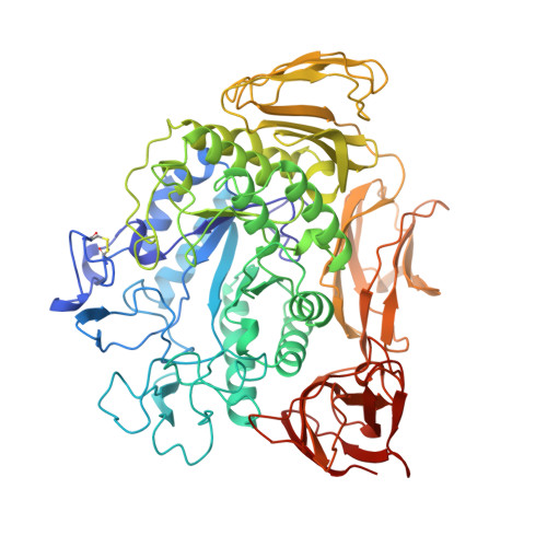
Structure of cyclodextrin glycosyltransferase complexed with a derivative of its main product beta-cyclodextrin.

Schmidt, A.K.Cottaz, S.Driguez, H.Schulz, G.E.

(1998) Biochemistry 37: 5909-5915

  • DOI: https://doi.org/10.1021/bi9729918
  • Primary Citation of Related Structures:  
    3CGT

  • PubMed Abstract: 

    Crystals of the inactive mutant Glu257-->Ala of cyclodextrin glycosyltransferase were soaked with the cyclodextrin (CD) derivative S-(alpha-D-glucopyranosyl)-6-thio-beta-CD. The structural analysis showed its beta-CD moiety with no density indication for the exocyclic glucosyl unit. For steric reasons, however, the position of this unit is restricted to be at only two of the seven glucosyl groups of beta-CD. The analysis indicated that the enzyme can cyclize branched alpha-glucans. The ligated beta-CD moiety revealed how the enzyme binds its predominant cyclic product. The conformation of the ligated beta-CD was intermediate between the more symmetrical conformation in beta-CD dodecahydrate crystals and the conformation of a bound linear alpha-glucan chain. Its scissile bond was displaced by 2.8 A from the position in linear alpha-glucans. Accordingly, the complex represents the situation after the cyclization reaction but before diffusion into the solvent, where a more symmetrical conformation is assumed, or the equivalent state in the reverse reaction. Furthermore, a unifying nomenclature for oligosaccharide-binding subsites in proteins is proposed.


  • Organizational Affiliation

    Institut für Organische Chemie und Biochemie, Albert-Ludwigs-Universität, Freiburg im Breisgau, Germany.


Macromolecules
Find similar proteins by:  (by identity cutoff)  |  3D Structure
Entity ID: 1
MoleculeChains Sequence LengthOrganismDetailsImage
CYCLODEXTRIN GLYCOSYLTRANSFERASE684Niallia circulansMutation(s): 1 
EC: 2.4.1.19
UniProt
Find proteins for P30920 (Niallia circulans)
Explore P30920 
Go to UniProtKB:  P30920
Entity Groups  
Sequence Clusters30% Identity50% Identity70% Identity90% Identity95% Identity100% Identity
UniProt GroupP30920
Sequence Annotations
Expand
  • Reference Sequence
Oligosaccharides

Help

Entity ID: 2
MoleculeChains Length2D Diagram Glycosylation3D Interactions
Cycloheptakis-(1-4)-(alpha-D-glucopyranose)
B
7N/A
Glycosylation Resources
GlyTouCan:  G01435GL
GlyCosmos:  G01435GL
Biologically Interesting Molecules (External Reference) 1 Unique
Experimental Data & Validation

Experimental Data

  • Method: X-RAY DIFFRACTION
  • Resolution: 2.40 Å
  • R-Value Free: 0.228 
  • R-Value Work: 0.177 
  • R-Value Observed: 0.177 
  • Space Group: P 21 21 21
Unit Cell:
Length ( Å )Angle ( ˚ )
a = 94.5α = 90
b = 105β = 90
c = 113.8γ = 90
Software Package:
Software NamePurpose
XDSdata scaling
XSCALEdata scaling
X-PLORmodel building
X-PLORrefinement
XDSdata reduction
X-PLORphasing

Structure Validation

View Full Validation Report


Entry History 

Deposition Data

Revision History  (Full details and data files)

  • Version 1.0: 1998-05-27
    Type: Initial release
  • Version 1.1: 2008-03-25
    Changes: Version format compliance
  • Version 1.2: 2011-07-13
    Changes: Version format compliance
  • Version 2.0: 2020-07-29
    Type: Remediation
    Reason: Carbohydrate remediation
    Changes: Atomic model, Data collection, Derived calculations, Non-polymer description, Structure summary
  • Version 2.1: 2021-11-03
    Changes: Database references, Structure summary