3C75

Paracoccus versutus methylamine dehydrogenase in complex with amicyanin


Experimental Data Snapshot

  • Method: X-RAY DIFFRACTION
  • Resolution: 2.50 Å
  • R-Value Free: 0.283 
  • R-Value Work: 0.235 
  • R-Value Observed: 0.235 

wwPDB Validation   3D Report Full Report


This is version 1.1 of the entry. See complete history


Literature

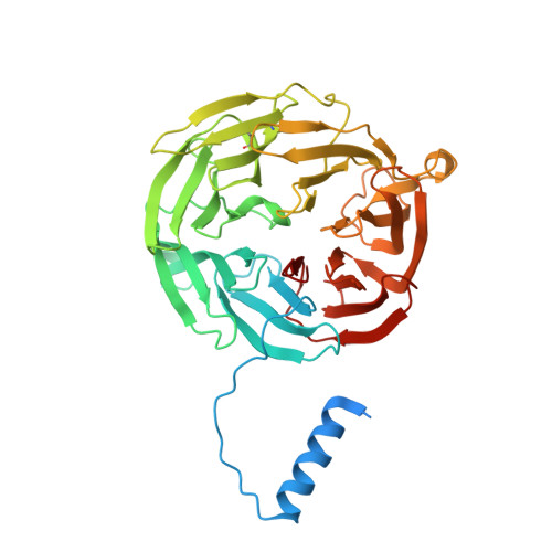
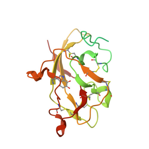
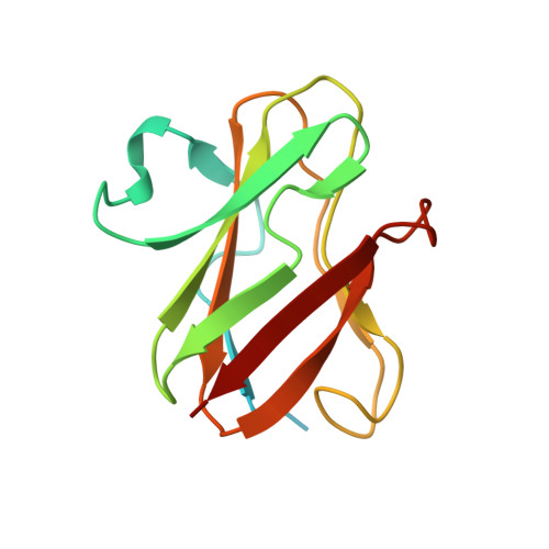
Structural comparison of crystal and solution states of the 138 kDa complex of methylamine dehydrogenase and amicyanin from Paracoccus versutus.

Cavalieri, C.Biermann, N.Vlasie, M.D.Einsle, O.Merli, A.Ferrari, D.Rossi, G.L.Ubbink, M.

(2008) Biochemistry 47: 6560-6570

  • DOI: https://doi.org/10.1021/bi7023749
  • Primary Citation of Related Structures:  
    3C75

  • PubMed Abstract: 

    Methylamine can be used as the sole carbon source of certain methylotrophic bacteria. Methylamine dehydrogenase catalyzes the conversion of methylamine into formaldehyde and donates electrons to the electron transfer protein amicyanin. The crystal structure of the complex of methylamine dehydrogenase and amicyanin from Paracoccus versutus has been determined, and the rate of electron transfer from the tryptophan tryptophylquinone cofactor of methylamine dehydrogenase to the copper ion of amicyanin in solution has been determined. In the presence of monovalent ions, the rate of electron transfer from the methylamine-reduced TTQ is much higher than in their absence. In general, the kinetics are similar to those observed for the system from Paracoccus denitrificans. The complex in solution has been studied using nuclear magnetic resonance. Signals of perdeuterated, (15)N-enriched amicyanin bound to methylamine dehydrogenase are observed. Chemical shift perturbation analysis indicates that the dissociation rate constant is approximately 250 s(-1) and that amicyanin assumes a well-defined position in the complex in solution. The most affected residues are in the interface observed in the crystal structure, whereas smaller chemical shift changes extend to deep inside the protein. These perturbations can be correlated to small differences in the hydrogen bond network observed in the crystal structures of free and bound amicyanin. This study indicates that chemical shift changes can be used as reliable indicators of subtle structural changes even in a complex larger than 100 kDa.


  • Organizational Affiliation

    Department of Biochemistry and Molecular Biology, University of Parma, 43100 Parma, Italy.


Macromolecules
Find similar proteins by:  (by identity cutoff)  |  3D Structure
Entity ID: 1
MoleculeChains Sequence LengthOrganismDetailsImage
Methylamine dehydrogenase heavy chainA [auth H],
D [auth J]
426Paracoccus versutusMutation(s): 0 
EC: 1.4.99.3
UniProt
Find proteins for P23006 (Paracoccus versutus)
Explore P23006 
Go to UniProtKB:  P23006
Entity Groups  
Sequence Clusters30% Identity50% Identity70% Identity90% Identity95% Identity100% Identity
UniProt GroupP23006
Sequence Annotations
Expand
  • Reference Sequence
Find similar proteins by:  (by identity cutoff)  |  3D Structure
Entity ID: 2
MoleculeChains Sequence LengthOrganismDetailsImage
Methylamine dehydrogenase light chainB [auth L],
E [auth M]
188Paracoccus versutusMutation(s): 0 
EC: 1.4.99.3
UniProt
Find proteins for P22641 (Paracoccus versutus)
Explore P22641 
Go to UniProtKB:  P22641
Entity Groups  
Sequence Clusters30% Identity50% Identity70% Identity90% Identity95% Identity100% Identity
UniProt GroupP22641
Sequence Annotations
Expand
  • Reference Sequence
Find similar proteins by:  (by identity cutoff)  |  3D Structure
Entity ID: 3
MoleculeChains Sequence LengthOrganismDetailsImage
AmicyaninC [auth A],
F [auth B]
132Paracoccus versutusMutation(s): 0 
UniProt
Find proteins for P22365 (Paracoccus versutus)
Explore P22365 
Go to UniProtKB:  P22365
Entity Groups  
Sequence Clusters30% Identity50% Identity70% Identity90% Identity95% Identity100% Identity
UniProt GroupP22365
Sequence Annotations
Expand
  • Reference Sequence
Small Molecules
Modified Residues  1 Unique
IDChains TypeFormula2D DiagramParent
TRQ
Query on TRQ
B [auth L],
E [auth M]
L-PEPTIDE LINKINGC11 H10 N2 O4TRP
Experimental Data & Validation

Experimental Data

  • Method: X-RAY DIFFRACTION
  • Resolution: 2.50 Å
  • R-Value Free: 0.283 
  • R-Value Work: 0.235 
  • R-Value Observed: 0.235 
  • Space Group: P 21 21 21
Unit Cell:
Length ( Å )Angle ( ˚ )
a = 55.573α = 90
b = 131.038β = 90
c = 171.689γ = 90
Software Package:
Software NamePurpose
MAR345dtbdata collection
AMoREphasing
CNSrefinement
DENZOdata reduction
SCALEPACKdata scaling

Structure Validation

View Full Validation Report


Entry History 

Deposition Data

Revision History  (Full details and data files)

  • Version 1.0: 2008-12-30
    Type: Initial release
  • Version 1.1: 2011-07-13
    Changes: Version format compliance