3B9A

Crystal structure of Vibrio harveyi chitinase A complexed with hexasaccharide


Experimental Data Snapshot

  • Method: X-RAY DIFFRACTION
  • Resolution: 1.80 Å
  • R-Value Free: 0.210 
  • R-Value Work: 0.181 
  • R-Value Observed: 0.182 

wwPDB Validation   3D Report Full Report


This is version 2.2 of the entry. See complete history


Literature

Crystal structures of Vibrio harveyi chitinase A complexed with chitooligosaccharides: implications for the catalytic mechanism

Songsiriritthigul, C.Pantoom, S.Aguda, A.H.Robinson, R.C.Suginta, W.

(2008) J Struct Biol 162: 491-499

  • DOI: https://doi.org/10.1016/j.jsb.2008.03.008
  • Primary Citation of Related Structures:  
    3B8S, 3B9A, 3B9D, 3B9E

  • PubMed Abstract: 

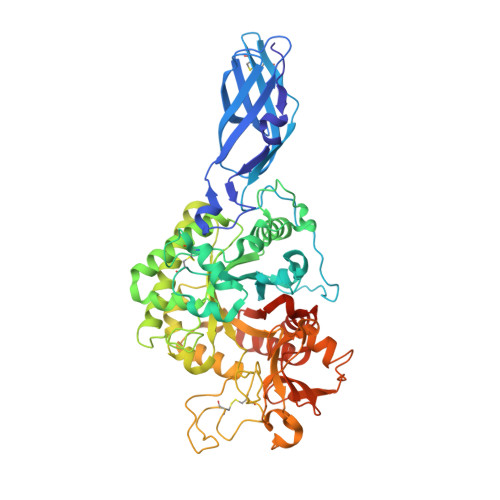
    This research describes four X-ray structures of Vibrio harveyi chitinase A and its catalytically inactive mutant (E315M) in the presence and absence of substrates. The overall structure of chitinase A is that of a typical family-18 glycosyl hydrolase comprising three distinct domains: (i) the amino-terminal chitin-binding domain; (ii) the main catalytic (alpha/beta)(8) TIM-barrel domain; and (iii) the small (alpha+beta) insertion domain. The catalytic cleft of chitinase A has a long, deep groove, which contains six chitooligosaccharide ring-binding subsites (-4)(-3)(-2)(-1)(+1)(+2). The binding cleft of the ligand-free E315M is partially blocked by the C-terminal (His)(6)-tag. Structures of E315M-chitooligosaccharide complexes display a linear conformation of pentaNAG, but a bent conformation of hexaNAG. Analysis of the final 2F(o)-F(c) omit map of E315M-NAG6 reveals the existence of the linear conformation of the hexaNAG at a lower occupancy with respect to the bent conformation. These crystallographic data provide evidence that the interacting sugars undergo conformational changes prior to hydrolysis by the wild-type enzyme.


  • Organizational Affiliation

    School of Biochemistry, Suranaree University of Technology, Nakhon Ratchasima 30000, Thailand.


Macromolecules
Find similar proteins by:  (by identity cutoff)  |  3D Structure
Entity ID: 1
MoleculeChains Sequence LengthOrganismDetailsImage
Chitinase A584Vibrio harveyiMutation(s): 1 
Gene Names: chiA
EC: 3.2.1.14
UniProt
Find proteins for Q9AMP1 (Vibrio harveyi)
Explore Q9AMP1 
Go to UniProtKB:  Q9AMP1
Entity Groups  
Sequence Clusters30% Identity50% Identity70% Identity90% Identity95% Identity100% Identity
UniProt GroupQ9AMP1
Sequence Annotations
Expand
  • Reference Sequence
Oligosaccharides

Help

Entity ID: 2
MoleculeChains Length2D Diagram Glycosylation3D Interactions
2-acetamido-2-deoxy-beta-D-glucopyranose-(1-4)-2-acetamido-2-deoxy-beta-D-glucopyranose-(1-4)-2-acetamido-2-deoxy-beta-D-glucopyranose-(1-4)-2-acetamido-2-deoxy-beta-D-glucopyranose-(1-4)-2-acetamido-2-deoxy-beta-D-glucopyranose-(1-4)-2-acetamido-2-deoxy-beta-D-glucopyranose
B
6N/A
Glycosylation Resources
GlyTouCan:  G36414FS
GlyCosmos:  G36414FS
GlyGen:  G36414FS
Experimental Data & Validation

Experimental Data

  • Method: X-RAY DIFFRACTION
  • Resolution: 1.80 Å
  • R-Value Free: 0.210 
  • R-Value Work: 0.181 
  • R-Value Observed: 0.182 
  • Space Group: P 21 21 21
Unit Cell:
Length ( Å )Angle ( ˚ )
a = 63.704α = 90
b = 83.323β = 90
c = 106.565γ = 90
Software Package:
Software NamePurpose
REFMACrefinement
CrystalCleardata collection
MOSFLMdata reduction
SCALAdata scaling
AMoREphasing

Structure Validation

View Full Validation Report


Entry History 

Deposition Data

Revision History  (Full details and data files)

  • Version 1.0: 2008-04-01
    Type: Initial release
  • Version 1.1: 2011-07-13
    Changes: Non-polymer description, Version format compliance
  • Version 2.0: 2020-07-29
    Type: Remediation
    Reason: Carbohydrate remediation
    Changes: Atomic model, Data collection, Database references, Derived calculations, Structure summary
  • Version 2.1: 2021-11-10
    Changes: Database references, Structure summary
  • Version 2.2: 2023-11-01
    Changes: Data collection, Refinement description