2V6E

protelomerase TelK complexed with substrate DNA


Experimental Data Snapshot

  • Method: X-RAY DIFFRACTION
  • Resolution: 3.20 Å
  • R-Value Free: 0.286 
  • R-Value Work: 0.255 
  • R-Value Observed: 0.255 

wwPDB Validation   3D Report Full Report


This is version 1.2 of the entry. See complete history


Literature

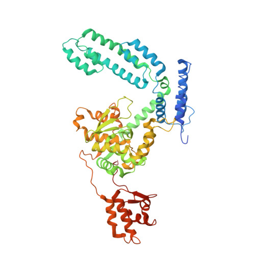
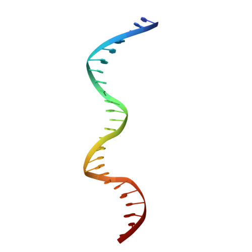
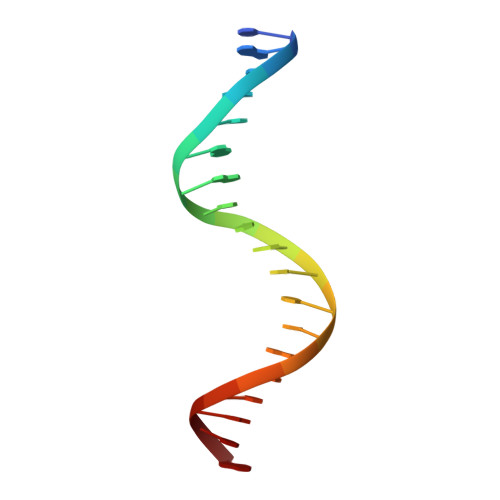
An Interlocked Dimer of the Protelomerase Telk Distorts DNA Structure for the Formation of Hairpin Telomeres

Aihara, H.Huang, W.M.Ellenberger, T.

(2007) Mol Cell 27: 901

  • DOI: https://doi.org/10.1016/j.molcel.2007.07.026
  • Primary Citation of Related Structures:  
    2V6E

  • PubMed Abstract: 

    The termini of linear chromosomes are protected by specialized DNA structures known as telomeres that also facilitate the complete replication of DNA ends. The simplest type of telomere is a covalently closed DNA hairpin structure found in linear chromosomes of prokaryotes and viruses. Bidirectional replication of a chromosome with hairpin telomeres produces a catenated circular dimer that is subsequently resolved into unit-length chromosomes by a dedicated DNA cleavage-rejoining enzyme known as a hairpin telomere resolvase (protelomerase). Here we report a crystal structure of the protelomerase TelK from Klebsiella oxytoca phage varphiKO2, in complex with the palindromic target DNA. The structure shows the TelK dimer destabilizes base pairing interactions to promote the refolding of cleaved DNA ends into two hairpin ends. We propose that the hairpinning reaction is made effectively irreversible by a unique protein-induced distortion of the DNA substrate that prevents religation of the cleaved DNA substrate.


  • Organizational Affiliation

    Department of Biochemistry and Molecular Biophysics, Washington University School of Medicine, 660 South Euclid Avenue, St. Louis, MO 63110, USA.


Macromolecules

Find similar proteins by:  (by identity cutoff)  |  3D Structure
Entity ID: 1
MoleculeChains Sequence LengthOrganismDetailsImage
PROTELEMORASE
A, B
558Klebsiella phage phiKO2Mutation(s): 0 
UniProt
Find proteins for Q6UAV6 (Klebsiella oxytoca phage phiKO2)
Explore Q6UAV6 
Go to UniProtKB:  Q6UAV6
Entity Groups  
Sequence Clusters30% Identity50% Identity70% Identity90% Identity95% Identity100% Identity
UniProt GroupQ6UAV6
Sequence Annotations
Expand
  • Reference Sequence

Find similar nucleic acids by:  Sequence   |   3D Structure  

Entity ID: 2
MoleculeChains LengthOrganismImage
TELRL
C, E
25Ravinvirus N15
Sequence Annotations
Expand
  • Reference Sequence

Find similar nucleic acids by:  Sequence   |   3D Structure  

Entity ID: 3
MoleculeChains LengthOrganismImage
TELRL
D, F
19Ravinvirus N15
Sequence Annotations
Expand
  • Reference Sequence
Experimental Data & Validation

Experimental Data

  • Method: X-RAY DIFFRACTION
  • Resolution: 3.20 Å
  • R-Value Free: 0.286 
  • R-Value Work: 0.255 
  • R-Value Observed: 0.255 
  • Space Group: P 41
Unit Cell:
Length ( Å )Angle ( ˚ )
a = 157.973α = 90
b = 157.973β = 90
c = 90.842γ = 90
Software Package:
Software NamePurpose
CNSrefinement
HKL-2000data reduction
HKL-2000data scaling
SOLVEphasing

Structure Validation

View Full Validation Report


Entry History 

Deposition Data

Revision History  (Full details and data files)

  • Version 1.0: 2007-10-02
    Type: Initial release
  • Version 1.1: 2011-05-08
    Changes: Version format compliance
  • Version 1.2: 2011-07-13
    Changes: Version format compliance