2BZZ

Crystal Structures of Eosinophil-derived Neurotoxin in Complex with the Inhibitors 5'-ATP, Ap3A, Ap4A and Ap5A


Experimental Data Snapshot

  • Method: X-RAY DIFFRACTION
  • Resolution: 0.98 Å

wwPDB Validation   3D Report Full Report


This is version 1.4 of the entry. See complete history


Literature

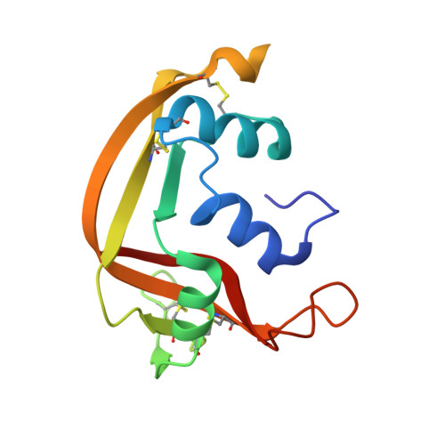
Crystal Structures of Eosinophil-Derived Neurotoxin (Edn) in Complex with the Inhibitors 5'- ATP, Ap(3)A, Ap(4)A, and Ap(5)A.

Baker, M.D.Holloway, D.E.Swaminathan, G.J.Acharya, K.R.

(2006) Biochemistry 45: 416

  • DOI: https://doi.org/10.1021/bi0518592
  • Primary Citation of Related Structures:  
    2BZZ, 2C01, 2C02, 2C05

  • PubMed Abstract: 

    Eosinophil-derived neurotoxin (EDN) is a catalytically proficient member of the pancreatic ribonuclease superfamily secreted along with other eosinophil granule proteins during innate host defense responses and various eosinophil-related inflammatory and allergic diseases. The ribonucleolytic activity of EDN is central to its antiviral and neurotoxic activities and possibly to other facets of its biological activity. To probe the importance of this enzymatic activity further, specific inhibitors will be of great aid. Derivatives of 5'-ADP are among the most potent inhibitors currently known. Here, we use X-ray crystallography to investigate the binding of four natural nucleotides containing this moiety. 5'-ATP binds in two alternative orientations, one occupying the B2 subsite in a conventional manner and one being a retro orientation with no ordered adenosine moiety. Diadenosine triphosphate (Ap3A) and diadenosine tetraphosphate (Ap4A) bind with one adenine positioned at the B2 subsite, the polyphosphate chain extending across the P1 subsite in an ill-defined conformation, and a disordered second adenosine moiety. Diadenosine pentaphosphate (Ap5A), the most avid inhibitor of this series, binds in a completely ordered fashion with one adenine positioned conventionally at the B2 subsite, the polyphosphate chain occupying the P1 and putative P(-1) subsites, and the other adenine bound in a retro-like manner at the edge of the B1 subsite. The binding mode of each of these inhibitors has features seen in previously determined structures of adenosine diphosphates. We examine the structure-affinity relationships of these inhibitors and discuss the implications for the design of improved inhibitors.


  • Organizational Affiliation

    Department of Biology and Biochemistry, University of Bath, Claverton Down, Bath BA2 7AY, United Kingdom.


Macromolecules
Find similar proteins by:  (by identity cutoff)  |  3D Structure
Entity ID: 1
MoleculeChains Sequence LengthOrganismDetailsImage
NONSECRETORY RIBONUCLEASE135Homo sapiensMutation(s): 0 
EC: 3.1.27.5
UniProt & NIH Common Fund Data Resources
Find proteins for P10153 (Homo sapiens)
Explore P10153 
Go to UniProtKB:  P10153
PHAROS:  P10153
GTEx:  ENSG00000169385 
Entity Groups  
Sequence Clusters30% Identity50% Identity70% Identity90% Identity95% Identity100% Identity
UniProt GroupP10153
Sequence Annotations
Expand
  • Reference Sequence
Small Molecules
Ligands 2 Unique
IDChains Name / Formula / InChI Key2D Diagram3D Interactions
AP5
Query on AP5

Download Ideal Coordinates CCD File 
B [auth A]BIS(ADENOSINE)-5'-PENTAPHOSPHATE
C20 H29 N10 O22 P5
OIMACDRJUANHTJ-XPWFQUROSA-N
ACY
Query on ACY

Download Ideal Coordinates CCD File 
C [auth A]ACETIC ACID
C2 H4 O2
QTBSBXVTEAMEQO-UHFFFAOYSA-N
Binding Affinity Annotations 
IDSourceBinding Affinity
AP5 Binding MOAD:  2BZZ Ki: 370 (nM) from 1 assay(s)
PDBBind:  2BZZ Ki: 370 (nM) from 1 assay(s)
Experimental Data & Validation

Experimental Data

  • Method: X-RAY DIFFRACTION
  • Resolution: 0.98 Å
  • Space Group: P 21 21 21
Unit Cell:
Length ( Å )Angle ( ˚ )
a = 53.036α = 90
b = 56.965β = 90
c = 42.259γ = 90
Software Package:
Software NamePurpose
SHELXL-97refinement
DENZOdata reduction
SCALEPACKdata scaling
AMoREphasing

Structure Validation

View Full Validation Report


Entry History 

Deposition Data

Revision History  (Full details and data files)

  • Version 1.0: 2006-01-18
    Type: Initial release
  • Version 1.1: 2011-05-08
    Changes: Version format compliance
  • Version 1.2: 2011-07-13
    Changes: Version format compliance
  • Version 1.3: 2019-10-16
    Changes: Advisory, Data collection, Experimental preparation, Other
  • Version 1.4: 2023-12-13
    Changes: Advisory, Data collection, Database references, Refinement description