1ZSO

Hypothetical protein from plasmodium falciparum


Experimental Data Snapshot

  • Method: X-RAY DIFFRACTION
  • Resolution: 2.17 Å
  • R-Value Free: 0.233 
  • R-Value Work: 0.183 
  • R-Value Observed: 0.186 

wwPDB Validation   3D Report Full Report


This is version 1.4 of the entry. See complete history


Literature

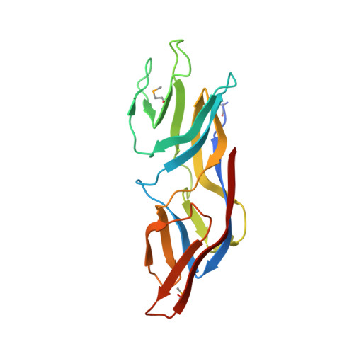
Structure of the conserved hypothetical protein MAL13P1.257 from Plasmodium falciparum.

Holmes, M.A.Buckner, F.S.Van Voorhis, W.C.Mehlin, C.Boni, E.Earnest, T.N.DeTitta, G.Luft, J.Lauricella, A.Anderson, L.Kalyuzhniy, O.Zucker, F.Schoenfeld, L.W.Hol, W.G.Merritt, E.A.

(2006) Acta Crystallogr Sect F Struct Biol Cryst Commun 62: 180-185

  • DOI: https://doi.org/10.1107/S1744309106005847
  • Primary Citation of Related Structures:  
    1ZSO

  • PubMed Abstract: 

    The structure of a conserved hypothetical protein, PlasmoDB sequence MAL13P1.257 from Plasmodium falciparum, Pfam sequence family PF05907, has been determined as part of the structural genomics effort of the Structural Genomics of Pathogenic Protozoa consortium. The structure was determined by multiple-wavelength anomalous dispersion at 2.17 A resolution. The structure is almost entirely beta-sheet; it consists of 15 beta-strands and one short 3(10)-helix and represents a new protein fold. The packing of the two monomers in the asymmetric unit indicates that the biological unit may be a dimer.


  • Organizational Affiliation

    Structural Genomics of Pathogenic Protozoa Consortium, USA.


Macromolecules
Find similar proteins by:  (by identity cutoff)  |  3D Structure
Entity ID: 1
MoleculeChains Sequence LengthOrganismDetailsImage
hypothetical protein
A, B
164Plasmodium falciparumMutation(s): 3 
Gene Names: MAL13P1.257
UniProt
Find proteins for Q8IDI8 (Plasmodium falciparum (isolate 3D7))
Explore Q8IDI8 
Go to UniProtKB:  Q8IDI8
Entity Groups  
Sequence Clusters30% Identity50% Identity70% Identity90% Identity95% Identity100% Identity
UniProt GroupQ8IDI8
Sequence Annotations
Expand
  • Reference Sequence
Small Molecules
Modified Residues  1 Unique
IDChains TypeFormula2D DiagramParent
MSE
Query on MSE
A, B
L-PEPTIDE LINKINGC5 H11 N O2 SeMET
Experimental Data & Validation

Experimental Data

  • Method: X-RAY DIFFRACTION
  • Resolution: 2.17 Å
  • R-Value Free: 0.233 
  • R-Value Work: 0.183 
  • R-Value Observed: 0.186 
  • Space Group: P 21 21 21
Unit Cell:
Length ( Å )Angle ( ˚ )
a = 62.247α = 90
b = 71.104β = 90
c = 78.616γ = 90
Software Package:
Software NamePurpose
SOLVEphasing
PDB_EXTRACTdata extraction
RESOLVEphasing
DENZOdata reduction
SCALEPACKdata scaling
REFMACrefinement

Structure Validation

View Full Validation Report


Entry History 

Deposition Data

Revision History  (Full details and data files)

  • Version 1.0: 2005-06-07
    Type: Initial release
  • Version 1.1: 2008-04-30
    Changes: Version format compliance
  • Version 1.2: 2011-07-13
    Changes: Advisory, Version format compliance
  • Version 1.3: 2012-10-24
    Changes: Database references
  • Version 1.4: 2017-10-11
    Changes: Refinement description