1OZ7

Crystal structure of Echicetin from the venom of Indian saw-scaled viper (Echis carinatus) at 2.4 resolution


Experimental Data Snapshot

  • Method: X-RAY DIFFRACTION
  • Resolution: 2.40 Å
  • R-Value Free: 0.244 
  • R-Value Work: 0.185 
  • R-Value Observed: 0.188 

wwPDB Validation   3D Report Full Report


This is version 1.3 of the entry. See complete history


Literature

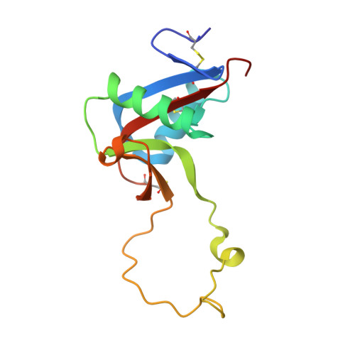
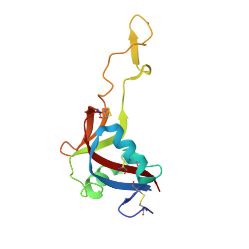
Crystal structure of echicetin from Echis carinatus (Indian saw-scaled viper) at 2.4A resolution.

Jasti, J.Paramasivam, M.Srinivasan, A.Singh, T.P.

(2004) J Mol Biol 335: 167-176

  • DOI: https://doi.org/10.1016/j.jmb.2003.10.048
  • Primary Citation of Related Structures:  
    1OZ7

  • PubMed Abstract: 

    Echicetin is a heterodimeric protein from the venom of the Indian saw-scaled viper, Echis carinatus. It binds to platelet glycoprotein Ib (GPIb) and thus inhibits platelet aggregation. It has two subunits, alpha and beta, consisting of 131 and 123 amino acid residues, respectively. The two chains are linked with a disulphide bond. The level of amino acid sequence homology between two subunits is 50%. The protein was purified from the venom of E.carinatus and crystallized using ammonium sulphate as a precipitant. The crystal structure has been determined at 2.4A resolution and refined to an R-factor of 0.187. Overall dimensions of the heterodimer are approximately 80Ax35Ax35A. The backbone folds of the two subunits are similar. The central portions of the polypeptide chains of alpha and beta-subunits move into each other to form a tight dimeric association. The remaining portions of the chains of both subunits fold in a manner similar to those observed in the carbohydrate-binding domains of C-type lectins. In echicetin, the Ca(2+)-binding sites are not present, despite being topologically equivalent to other similar Ca(2+)-binding proteins of the superfamily. The residues Ser41, Glu43 and Glu47 in the calcium-binding proteins of the related family are conserved but the residues Glu126/120 are replaced by lysine at the corresponding sites in the alpha and beta-subunits.


  • Organizational Affiliation

    Department of Biophysics, All India Institute of Medical Sciences, Ansari Nagar, 110 029, New Delhi, India.


Macromolecules
Find similar proteins by:  (by identity cutoff)  |  3D Structure
Entity ID: 1
MoleculeChains Sequence LengthOrganismDetailsImage
echicetin A-chain131Echis carinatusMutation(s): 0 
UniProt
Find proteins for Q7T248 (Echis carinatus)
Explore Q7T248 
Go to UniProtKB:  Q7T248
Entity Groups  
Sequence Clusters30% Identity50% Identity70% Identity90% Identity95% Identity100% Identity
UniProt GroupQ7T248
Sequence Annotations
Expand
  • Reference Sequence
Find similar proteins by:  (by identity cutoff)  |  3D Structure
Entity ID: 2
MoleculeChains Sequence LengthOrganismDetailsImage
echicetin B-chain123Echis carinatusMutation(s): 0 
UniProt
Find proteins for Q7T247 (Echis carinatus)
Explore Q7T247 
Go to UniProtKB:  Q7T247
Entity Groups  
Sequence Clusters30% Identity50% Identity70% Identity90% Identity95% Identity100% Identity
UniProt GroupQ7T247
Sequence Annotations
Expand
  • Reference Sequence
Experimental Data & Validation

Experimental Data

  • Method: X-RAY DIFFRACTION
  • Resolution: 2.40 Å
  • R-Value Free: 0.244 
  • R-Value Work: 0.185 
  • R-Value Observed: 0.188 
  • Space Group: P 1 21 1
Unit Cell:
Length ( Å )Angle ( ˚ )
a = 37.966α = 90
b = 95.258β = 112.58
c = 42.611γ = 90
Software Package:
Software NamePurpose
CNSrefinement
DENZOdata reduction
SCALEPACKdata scaling
AMoREphasing
REFMACrefinement

Structure Validation

View Full Validation Report


Entry History 

Deposition Data

Revision History  (Full details and data files)

  • Version 1.0: 2003-12-30
    Type: Initial release
  • Version 1.1: 2008-04-29
    Changes: Version format compliance
  • Version 1.2: 2011-07-13
    Changes: Version format compliance
  • Version 1.3: 2023-10-25
    Changes: Data collection, Database references, Refinement description