1MG3

MUTATION OF ALPHA PHE55 OF METHYLAMINE DEHYDROGENASE ALTERS THE REORGANIZATION ENERGY AND ELECTRONIC COUPLING FOR ITS ELECTRON TRANSFER REACTION WITH AMICYANIN


Experimental Data Snapshot

  • Method: X-RAY DIFFRACTION
  • Resolution: 2.40 Å
  • R-Value Free: 0.246 
  • R-Value Work: 0.193 

wwPDB Validation   3D Report Full Report


Ligand Structure Quality Assessment 


This is version 2.1 of the entry. See complete history


Literature

MUTATION OF ALPHA PHE55 OF METHYLAMINE DEHYDROGENASE ALTERS THE REORGANIZATION ENERGY AND ELECTRONIC COUPLING FOR ITS ELECTRON TRANSFER REACTION WITH AMICYANIN

Sun, D.Chen, Z.W.Mathews, F.S.Davidson, V.L.

(2002) Biochemistry 41: 13926-13933

  • DOI: https://doi.org/10.1021/bi026654x
  • Primary Citation of Related Structures:  
    1MG2, 1MG3

  • PubMed Abstract: 

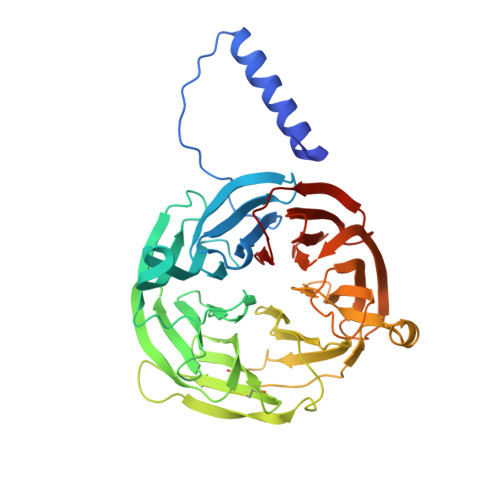
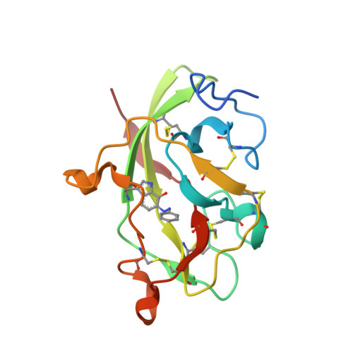
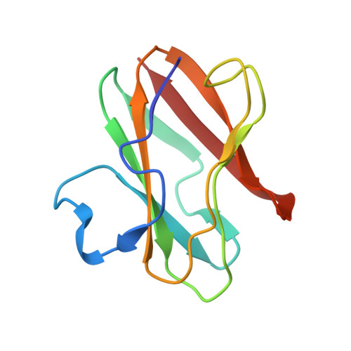
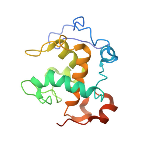
    Methylamine dehydrogenase (MADH) possesses an alpha(2)beta(2) structure with each smaller beta subunit possessing a tryptophan tryptophylquinone (TTQ) prosthetic group. Phe55 of the alpha subunit is located where the substrate channel from the enzyme surface opens into the active site. Site-directed mutagenesis of alphaPhe55 has revealed roles for this residue in determining substrate specificity and binding monovalent cations at the active site. It is now shown that the alphaF55A mutation also increases the rate of the true electron transfer (ET) reaction from O-quinol MADH to amicyanin. The reorganization energy associated with the ET reaction is decreased from 2.3 to 1.8 eV. The electronic coupling associated with the ET reaction is decreased from 12 to 3 cm(-1). The crystal structure of alphaF55A MADH in complex with its electron acceptors, amicyanin and cytochrome c-551i, has been determined. Little difference in the overall structure is seen, relative to the native complex; however, there are significant changes in the solvent content of the active site and substrate channel. The crystal structure of alphaF55A MADH has also been determined with phenylhydrazine covalently bound to TTQ in the active site. Phenylhydrazine binding significantly perturbs the orientation of the TTQ rings relative to each other. The ET results are discussed in the context of the new and old crystal structures of the native and mutant enzymes.


  • Organizational Affiliation

    Department of Biochemistry, The University of Mississippi Medical Center, Jackson, Mississippi 39216-4505, USA.


Macromolecules
Find similar proteins by:  (by identity cutoff)  |  3D Structure
Entity ID: 1
MoleculeChains Sequence LengthOrganismDetailsImage
Methylamine dehydrogenase, heavy chain
A, E, I, M
390Paracoccus denitrificansMutation(s): 1 
EC: 1.4.99.3
UniProt
Find proteins for P29894 (Paracoccus denitrificans)
Explore P29894 
Go to UniProtKB:  P29894
Entity Groups  
Sequence Clusters30% Identity50% Identity70% Identity90% Identity95% Identity100% Identity
UniProt GroupP29894
Sequence Annotations
Expand
  • Reference Sequence
Find similar proteins by:  (by identity cutoff)  |  3D Structure
Entity ID: 2
MoleculeChains Sequence LengthOrganismDetailsImage
Methylamine dehydrogenase, light chain
B, F, J, N
131Paracoccus denitrificansMutation(s): 1 
EC: 1.4.99.3
UniProt
Find proteins for P22619 (Paracoccus denitrificans)
Explore P22619 
Go to UniProtKB:  P22619
Entity Groups  
Sequence Clusters30% Identity50% Identity70% Identity90% Identity95% Identity100% Identity
UniProt GroupP22619
Sequence Annotations
Expand
  • Reference Sequence
Find similar proteins by:  (by identity cutoff)  |  3D Structure
Entity ID: 3
MoleculeChains Sequence LengthOrganismDetailsImage
Amicyanin
C, G, K, O
105Paracoccus denitrificansMutation(s): 0 
UniProt
Find proteins for P22364 (Paracoccus denitrificans)
Explore P22364 
Go to UniProtKB:  P22364
Entity Groups  
Sequence Clusters30% Identity50% Identity70% Identity90% Identity95% Identity100% Identity
UniProt GroupP22364
Sequence Annotations
Expand
  • Reference Sequence
Find similar proteins by:  (by identity cutoff)  |  3D Structure
Entity ID: 4
MoleculeChains Sequence LengthOrganismDetailsImage
CYTOCHROME C-L
D, H, L, P
155Paracoccus denitrificansMutation(s): 0 
UniProt
Find proteins for P29899 (Paracoccus denitrificans)
Explore P29899 
Go to UniProtKB:  P29899
Entity Groups  
Sequence Clusters30% Identity50% Identity70% Identity90% Identity95% Identity100% Identity
UniProt GroupP29899
Sequence Annotations
Expand
  • Reference Sequence
Small Molecules
Ligands 4 Unique
IDChains Name / Formula / InChI Key2D Diagram3D Interactions
HEC
Query on HEC

Download Ideal Coordinates CCD File 
EA [auth L],
JA [auth P],
U [auth D],
Z [auth H]
HEME C
C34 H34 Fe N4 O4
HXQIYSLZKNYNMH-LJNAALQVSA-N
PO4
Query on PO4

Download Ideal Coordinates CCD File 
AA [auth I]
BA [auth J]
FA [auth M]
HA [auth O]
Q [auth A]
AA [auth I],
BA [auth J],
FA [auth M],
HA [auth O],
Q [auth A],
R [auth B],
V [auth E],
W [auth F]
PHOSPHATE ION
O4 P
NBIIXXVUZAFLBC-UHFFFAOYSA-K
CU
Query on CU

Download Ideal Coordinates CCD File 
CA [auth K],
GA [auth O],
S [auth C],
X [auth G]
COPPER (II) ION
Cu
JPVYNHNXODAKFH-UHFFFAOYSA-N
NA
Query on NA

Download Ideal Coordinates CCD File 
DA [auth L],
IA [auth P],
T [auth D],
Y [auth H]
SODIUM ION
Na
FKNQFGJONOIPTF-UHFFFAOYSA-N
Modified Residues  1 Unique
IDChains TypeFormula2D DiagramParent
TRW
Query on TRW
B, F, J, N
L-PEPTIDE LINKINGC17 H18 N4 O2TRP
Experimental Data & Validation

Experimental Data

  • Method: X-RAY DIFFRACTION
  • Resolution: 2.40 Å
  • R-Value Free: 0.246 
  • R-Value Work: 0.193 
  • Space Group: P 1 21 1
Unit Cell:
Length ( Å )Angle ( ˚ )
a = 79.52α = 90
b = 188.372β = 98.84
c = 127.366γ = 90
Software Package:
Software NamePurpose
SCALEPACKdata scaling
CNSrefinement

Structure Validation

View Full Validation Report


Ligand Structure Quality Assessment 


Entry History 

Deposition Data

Revision History  (Full details and data files)

  • Version 1.0: 2002-12-11
    Type: Initial release
  • Version 1.1: 2008-04-28
    Changes: Version format compliance
  • Version 1.2: 2011-07-13
    Changes: Version format compliance
  • Version 1.3: 2017-10-11
    Changes: Refinement description
  • Version 2.0: 2021-03-03
    Changes: Advisory, Atomic model, Data collection, Database references, Derived calculations, Non-polymer description, Structure summary
  • Version 2.1: 2021-10-27
    Changes: Database references