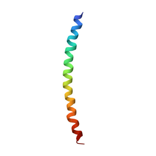
Structure of the capsid of Pf3 filamentous phage determined from X-ray fibre diffraction data at 3.1 A resolution.
Welsh, L.C., Symmons, M.F., Sturtevant, J.M., Marvin, D.A., Perham, R.N.(1998) J Mol Biology 283: 155-177
- PubMed: 9761681
- DOI: https://doi.org/10.1006/jmbi.1998.2081
- Primary Citation of Related Structures:
1IFP - PubMed Abstract:
We have recorded X-ray diffraction patterns at 3.1 A resolution from magnetically aligned fibres of the Pf3 strain of filamentous bacteriophage (Inovirus). The patterns are similar to patterns from the higher-temperature form of the Pf1 strain, indicating that the Pf3 and Pf1 virions have the same helix symmetry and similar protein subunit shape. This is of particular interest, given that the primary structures of the two protein subunits are quite different; and the nucleotide/protein subunit ratio in the Pf3 virion is more than twice that in Pf1, indicating important differences in DNA packaging. We have built a molecular model of the Pf3 protein capsid based on the model of Pf1, and refined it against the diffraction data using simulated annealing. The refinement confirms that the two structures are similar, which may reflect a fundamental motif of alpha-helix packing. However, there are some differences between the structures: the Pf3 subunit appears to be completely alpha-helical, beginning at the N terminus, whereas the first few residues of the Pf1 subunit are not helical; and the structure of the C-terminal region of the Pf3 subunit at the inner surface of the tubular capsid indicates that DNA/protein interactions in this virion may involve both aromatic side-chains and positively charged side-chains, whereas those in the Pf1 virion involve predominantly only the latter. In the course of this work, we have developed new approaches to refinement and validation of helical structures with respect to continuous transform fibre diffraction data.
- Department of Biochemistry, University of Cambridge, 80 Tennis Court Road, Cambridge, CB2 1GA, UK.
Organizational Affiliation: