1IAW

CRYSTAL STRUCTURE OF NAEI COMPLEXED WITH 17MER DNA


Experimental Data Snapshot

  • Method: X-RAY DIFFRACTION
  • Resolution: 2.40 Å
  • R-Value Free: 0.293 
  • R-Value Work: 0.243 

wwPDB Validation   3D Report Full Report


This is version 1.5 of the entry. See complete history


Literature

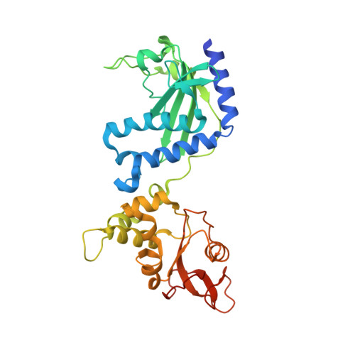
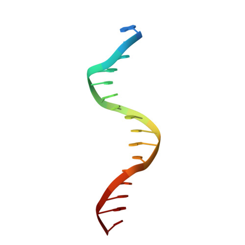
Structure of NaeI-DNA complex reveals dual-mode DNA recognition and complete dimer rearrangement.

Huai, Q.Colandene, J.D.Topal, M.D.Ke, H.

(2001) Nat Struct Biol 8: 665-669

  • DOI: https://doi.org/10.1038/90366
  • Primary Citation of Related Structures:  
    1IAW

  • PubMed Abstract: 

    NaeI, a novel DNA endonuclease, shows topoisomerase and recombinase activities when a Lys residue is substituted for Leu 43. The NaeI-DNA structure demonstrates that each of the two domains of NaeI recognizes one molecule of DNA duplex. DNA recognition induces dramatic rearrangements: narrowing the binding site of the Topo domain 16 A to grip DNA, widening that of the Endo domain 8 A to encircle and bend DNA 45 degrees for cleavage, and completely rebuilding the homodimer interface. The NaeI-DNA structure presents the first example of novel recognition of two copies of one DNA sequence by two different amino acid sequences and two different structural motifs in one polypeptide.


  • Organizational Affiliation

    Department of Biochemistry and Biophysics, The University of North Carolina, Chapel Hill, North Carolina 27599-7260, USA.


Macromolecules

Find similar proteins by:  (by identity cutoff)  |  3D Structure
Entity ID: 2
MoleculeChains Sequence LengthOrganismDetailsImage
TYPE II RESTRICTION ENZYME NAEIE [auth A],
F [auth B]
317Lentzea aerocolonigenesMutation(s): 0 
Gene Names: NAE I
EC: 3.1.21.4
UniProt
Find proteins for P50187 (Lentzea aerocolonigenes)
Explore P50187 
Go to UniProtKB:  P50187
Entity Groups  
Sequence Clusters30% Identity50% Identity70% Identity90% Identity95% Identity100% Identity
UniProt GroupP50187
Sequence Annotations
Expand
  • Reference Sequence

Find similar nucleic acids by:  Sequence   |   3D Structure  

Entity ID: 1
MoleculeChains LengthOrganismImage
5'-D(*TP*GP*CP*CP*AP*CP*GP*CP*CP*GP*GP*CP*GP*TP*GP*GP*C)-3'A [auth C],
B [auth D],
C [auth E],
D [auth F]
17N/A
Sequence Annotations
Expand
  • Reference Sequence
Experimental Data & Validation

Experimental Data

  • Method: X-RAY DIFFRACTION
  • Resolution: 2.40 Å
  • R-Value Free: 0.293 
  • R-Value Work: 0.243 
  • Space Group: P 21 21 21
Unit Cell:
Length ( Å )Angle ( ˚ )
a = 59.6α = 90
b = 65β = 90
c = 258.5γ = 90
Software Package:
Software NamePurpose
AMoREphasing
CNSrefinement
DENZOdata reduction
SCALEPACKdata scaling

Structure Validation

View Full Validation Report


Entry History 

Deposition Data

Revision History  (Full details and data files)

  • Version 1.0: 2001-08-03
    Type: Initial release
  • Version 1.1: 2008-04-27
    Changes: Version format compliance
  • Version 1.2: 2011-07-13
    Changes: Version format compliance
  • Version 1.3: 2012-07-25
    Changes: Other
  • Version 1.4: 2024-02-07
    Changes: Data collection, Database references
  • Version 1.5: 2024-04-03
    Changes: Refinement description