9ZD6 | pdb_00009zd6

Bacterial interstrand DNA crosslink glycosylase AlkX/YcaQ bound to DNA


Experimental Data Snapshot

  • Method: X-RAY DIFFRACTION
  • Resolution: 2.60 Å
  • R-Value Free: 
    0.256 (Depositor), 0.256 (DCC) 
  • R-Value Work: 
    0.207 (Depositor), 0.207 (DCC) 
  • R-Value Observed: 
    0.209 (Depositor) 

Starting Model: experimental
View more details

wwPDB Validation   3D Report Full Report


This is version 1.0 of the entry. See complete history


Literature

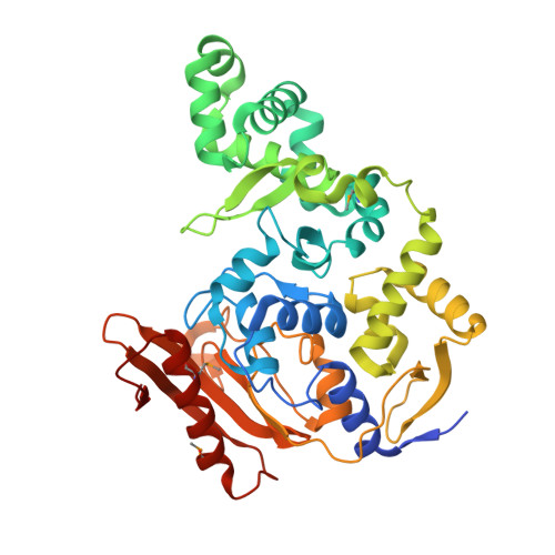
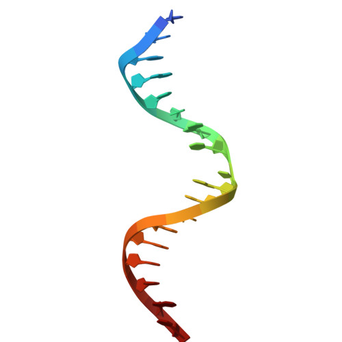
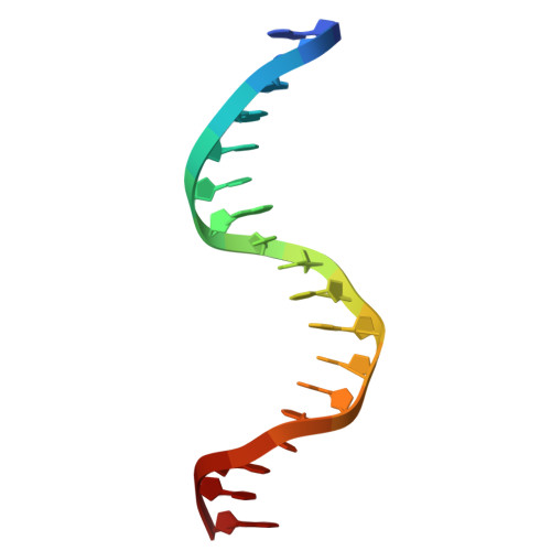
DNA binding and lesion recognition by the bacterial interstrand DNA crosslink glycosylase AlkX.

Cai 蔡毓娟, Y.Kunkle, D.E.Edinbugh, M.D.Skaar, E.P.Eichman, B.F.

(2025) bioRxiv 

  • DOI: https://doi.org/10.64898/2025.12.11.693820
  • Primary Citation of Related Structures:  
    9ZD6

  • PubMed Abstract: 

    Interstrand DNA crosslinks (ICLs) are a highly toxic form of DNA damage. ICL repair in both eukaryotes and bacteria involves unhooking of the two strands by specialized DNA glycosylases. We recently established that the human pathogen Acinetobacter baumannii contains an ICL glycosylase (AlkX) that facilitates pathogenesis and protects the bacteria from DNA damage and acid stress. However, the physical basis for glycosylase-catalyzed ICL unhooking is unknown. Here, we describe a crystal structure of AlkX bound to DNA representing a product of the ICL unhooking reaction. Mutational analysis of ICL unhooking in vitro and A. baumannii sensitivity to the crosslinking agent mechlorethamine enabled identification of several AlkX motifs critical for ICL repair. We also found that a genetic variant from an antibiotic-resistant strain of the human pathogen Salmonella enterica significantly reduced AlkX activity in vitro and increased A. baumannii sensitivity to DNA crosslinking. This work provides a structural basis for how bacterial ICL glycosylases recognize and repair DNA adducts and contributes additional evidence that ICL repair is important for fitness of human pathogens.


  • Organizational Affiliation
    • Department of Biological Sciences, Vanderbilt University, Nashville, Tennessee 37232.

Macromolecules

Find similar proteins by:  (by identity cutoff)  |  3D Structure
Entity ID: 1
MoleculeChains Sequence LengthOrganismDetailsImage
Cytoplasmic protein
A, B
402Thermobifida fuscaMutation(s): 0 
Gene Names: Tfu_0254
UniProt
Find proteins for Q47TC5 (Thermobifida fusca (strain YX))
Explore Q47TC5 
Go to UniProtKB:  Q47TC5
Entity Groups  
Sequence Clusters30% Identity50% Identity70% Identity90% Identity95% Identity100% Identity
UniProt GroupQ47TC5
Sequence Annotations
Expand
  • Reference Sequence

Find similar nucleic acids by:  Sequence   |   3D Structure  

Entity ID: 2
MoleculeChains LengthOrganismImage
DNA (5'-D(*TP*GP*AP*GP*TP*CP*GP*T*(3DR)P*GP*AP*TP*GP*AP*CP*CP*AP*C)-3')18DNA molecule
Sequence Annotations
Expand
  • Reference Sequence

Find similar nucleic acids by:  Sequence   |   3D Structure  

Entity ID: 3
MoleculeChains LengthOrganismImage
DNA (5'-D(*GP*TP*GP*GP*TP*CP*AP*TP*CP*CP*AP*CP*GP*AP*CP*TP*CP*A)-3')18DNA molecule
Sequence Annotations
Expand
  • Reference Sequence
Small Molecules
Modified Residues  1 Unique
IDChains TypeFormula2D DiagramParent
MSE
Query on MSE
A, B
L-PEPTIDE LINKINGC5 H11 N O2 SeMET
Experimental Data & Validation

Experimental Data

  • Method: X-RAY DIFFRACTION
  • Resolution: 2.60 Å
  • R-Value Free:  0.256 (Depositor), 0.256 (DCC) 
  • R-Value Work:  0.207 (Depositor), 0.207 (DCC) 
  • R-Value Observed: 0.209 (Depositor) 
Space Group: P 21 21 21
Unit Cell:
Length ( Å )Angle ( ˚ )
a = 56.598α = 90
b = 115.641β = 90
c = 160.676γ = 90
Software Package:
Software NamePurpose
PHENIXrefinement
Aimlessdata scaling
XDSdata reduction
PHENIXphasing
PDB_EXTRACTdata extraction

Structure Validation

View Full Validation Report


Entry History & Funding Information

Deposition Data


Funding OrganizationLocationGrant Number
National Science Foundation (NSF, United States)United States2341288

Revision History  (Full details and data files)

  • Version 1.0: 2026-01-14
    Type: Initial release