Download Mendeley
A bundled anti-parallel cytochrome nanowire structure suggests roles in cell-cell electron transfer and biofilm formation.
Petersen, H.A., Chan, C.H., Carpenter, G.O., Tabari, M.Z., Fields, J.L., Zia, A., Rich-New, S.T., Hochbaum, A.I., Bond, D.R., Wang, F.(2026) mBio : e0090626-e0090626
- PubMed: 42644614 Search on PubMed
- DOI: https://doi.org/10.1128/mbio.00906-26
- Primary Citation Related Structures:
9YUQ - PubMed Abstract:
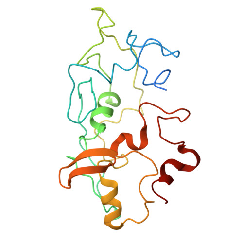
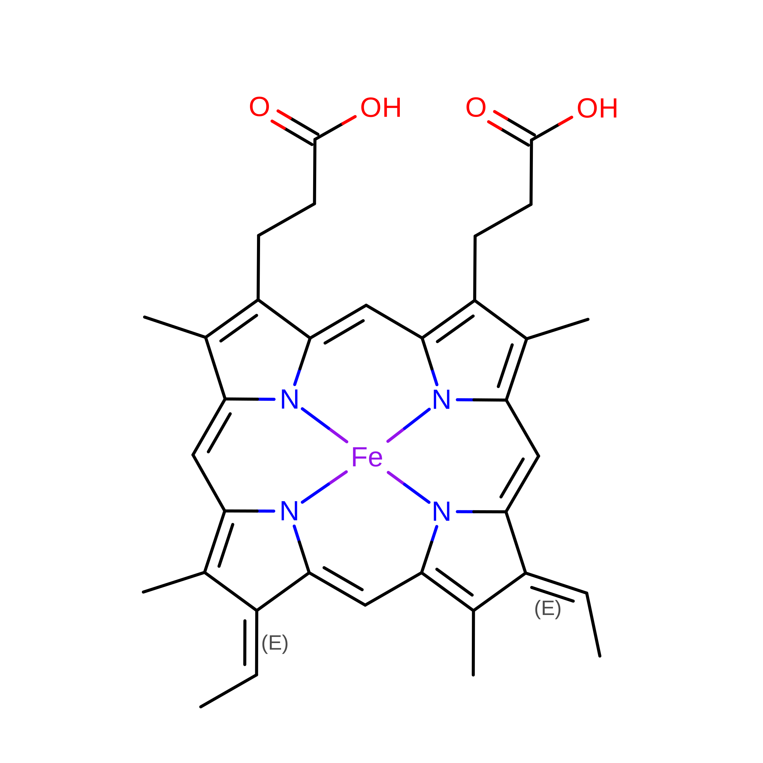
Long-range extracellular electron transfer enables respiring microbes to use minerals, organisms, or electrodes as electron acceptors by transporting electrons microns away from the cell surface. This process is primarily studied in Geobacter sulfurreducens , which produces at least three distinct, micrometer-long, multiheme cytochrome nanowires capable of electron transfer. However, the distribution and higher-order structure of such nanowires remains largely unknown. Here, we employed cryo-electron microscopy to determine the atomic structure of a unique cytochrome nanowire from Desulfuromonas soudanensis WTL, a halophilic, iron- and electrode-reducing bacterium from deep subsurface brine. These filaments are based on an OmcE tetraheme cytochrome homolog that assembles into highly ordered bundles of anti-parallel filaments. Electronic measurements of purified nanowire bundle films reveal that these bundles are conductive, with conductance increasing in a concentration-dependent manner. The apparent midpoint potential of these OmcE bundles was -145 mV vs SHE. They exhibit redox activity over a ~370 mV window attributable to five electrostatically coupled hemes, suggesting heme-heme interaction beyond the boundaries of individual OmcE repeat units. Observations of these bundles under diverse buffer conditions suggest that they represent stable supramolecular assemblies, although direct in vivo confirmation within intact biofilms remains a future goal. Furthermore, a similar cytochrome bundle structure was observed in Geobacter metallireducens , showing that this quaternary structure may be a common feature among nanowires secreted by electroactive microbes. Our findings demonstrate that cytochrome nanowires are widespread and can form specialized bundle interfaces. This novel state could facilitate conductive biofilm formation and form the basis for direct microbial electron exchange. IMPORTANCE All available structures of filaments containing a central core of closely spaced multiheme c-type cytochromes are derived from only a few organisms and are based on individual fibers easily imaged by cryo-electron microscopy. While imaging enriched preparations of extracellular appendages from a bacterium isolated from deep subsurface brine, we observed ordered bundles of anti-parallel cytochrome filaments. This quaternary arrangement contained specific contacts between adjacent nanowires, and a similar structure could be found in a relative from a related genus. This first report of bundling by nanowires suggests that such conductive structures may be missed in traditional surveys searching for novel filaments and provides an explanation for how nanowires could directly link adjacent cells in biofilms and granular communities.
- Department of Biochemistry and Molecular Genetics, University of Alabama at Birmingham, Birmingham, Alabama, USA.
Organizational Affiliation: